512 9 Management of foot and ankle disorders CHAPTER CONTENTS Introduction �������������������������������������������������������� 512 Musculoskeleta...
8 downloads
21 Views
5MB Size
Management of foot and ankle disorders
9
Jukka Kangas
CHAPTER CONTENTS Introduction �������������������������������������������������������� 512 Musculoskeletal foot and ankle disorders�������� 516 Psychosocial factors of pain and disability������ 518 Lifestyle factors and musculoskeletal foot � and ankle disorders�������������������������������������������� 519 Work-related factors and musculoskeletal � foot and ankle disorders������������������������������������ 519 Subjective examination�������������������������������������� 520 Planning the physical examination�������������������� 523 Physical examination������������������������������������������ 525 Treatment techniques ���������������������������������������� 528
Key words Anatomy and biomechanics, classification, clinical practice framework, examination, interventions, motor control impairment, movement behaviour, movement impairment, therapeutic exercise, passive movement
Introduction Foot and ankle problems are highly prevalent. Population-based studies indicate that between 18 and 63% of people report pain, aching, or stiffness in their feet (Hill et al. 2008, Menz et al. 2010). Musculoskeletal foot and ankle problems are commonly seen in primary care consisting of 8% of all musculoskeletal consultations (Menz et al. 2010). 512
The role of manual therapy in the intervention of musculoskeletal foot and ankle disorders is well established (Bronfort et al. 2010). In contemporary manual therapy practice musculoskeletal foot and ankle disorders should be considered in a multifactorial bio-psychosocial framework (Kangas et al. 2011). Therefore, the role of manual therapy extends far beyond manual techniques. Manual therapy practice should be a process involving the evaluation of the disorder and implementing an intervention based on evaluation. This process should consider the physical and the psychosocial dimensions of the disorder.
Anatomy and regions of the foot and ankle The foot consists of 28 irregularly shaped bones, over 30 joints, 32 muscles and over 100 ligaments. All these structures have to work synchronously to fulfill the high requirements of the foot and ankle. The foot alternates in form and function between a flexible structure for shock-absorption during loading response and a rigid lever for propulsion during terminal stance of the gait cycle. Considerable forces act on the foot and ankle during normal daily activities. For example, a joint contact force for the talocrural joint (TCJ) can range from three to five times the body weight during the stance phase of gait (Kleipool & Blankevoort 2010). The foot can be divided into different regions based on anatomy, arches of the feet, functional regions and for examination and treatment purposes.
Introduction
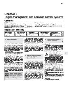
Anatomically the foot can be divided into hindfoot, midfoot, and forefoot (Fig. 9.1). The hindfoot consists of the talus and the calcaneus, the midfoot consists of the navicular, cuboid and cuneiform bones and the forefoot consists of the metatarsals (referred to as rays) and phalanges (Hamill et al. 1995). Classically, the arches of the foot have been defined as medial longitudinal, lateral longitudinal and transverse. From a functional perspective, the arches can be divided into medial, central and lateral
(Fig. 9.2). The medial arch consists of the talus, calcaneus, navicular, medial cuneiform and the first metatarsal bones. The first metatarsal, medial cuneiform and the navicular bones form the first ray. The central arch consists of the intermediate and lateral cuneiforms and the second and the third metatarsal bones. The lateral arch consists of the calcaneus, cuboid and the fourth and the fifth metatarsal bones. The fifth metatarsal and cuboid bones form the fifth ray.
The bones and arches of the foot
A
B
Talus Calcaneus Metatarsal Cuneiform Navicular I–V Cuboid
C
D
Figure 9.1 • The bones of the foot: A dorsal aspect; B plantar aspect; C lateral aspect with the lateral longitudinal arch; D medial aspect with the medial longitudinal arch. Figure 9.2 • Medial, central and lateral arches of the foot. The shaded area shows the central arch of the foot.
513
CHAPTER 9
Management of foot and ankle disorders
The medial and lateral arches are more flexible and are actively weight-bearing arches. Whereas, the central arch is more unyielding and passively contributes to the foot and ankle construct. For manual examination and treatment it is convenient to divide the foot and ankle into the hindfoot and forefoot. The hindfoot consists of the distal tibiofibular joint, talocrural joint (TCJ), subtalar joint (STJ) and surrounding soft tissue structures. The forefoot consists of the midtarsal joint (MTJ), intertarsal joints, tarsometatarsal (TMT) joints, intermetatarsal spaces, metatarsophalangeal (MTP) joints, interphalangeal (IP) joints, and surrounding soft tissue structures.
Movements of the foot and ankle Optimal function of the foot and ankle is based on the synchronization of the movements of individual joints. Single-joint movements occur in distinctively different directions (Nester et al. 2001, Arndt et al. 2004, Tweed et al. 2008). However, in functional movements of the foot and ankle independent movements of single joints are seldom, if impossible. The actions of the joints are highly inter-related and an action at one single joint will influence the other joints. Moreover, movements are often combined or coupled between the joints. For example, in the ankle one-third of the inversion and eversion occurs in TCJ and two-thirds in STJ (Kleipool & Blankevoort 2010).
Axes and planes of movements In this text, the terms eversion and inversion (EV and INV, respectively) are used to describe movements around an anteroposterior axis of the foot. These movements take place in the frontal plane. Plantar flexion and dorsiflexion are used to describe movements around a transverse axis of the foot. These movements take place in the sagittal plane. Abduction and adduction are used to describe movements around a vertical axis of the foot. These movements take place in the transverse plane (Arndt et al. 2004). The terms pronation and supination (Pron and Sup) are used to describe the triplanar motions of the subtalar joint (STJ) and midtarsal joint (MTJ). These movements are assumed to occur around an axis of the STJ and MTJ (Nester et al. 2001, Arndt et al. 2004) These definitions are used throughout the text – apart from in the section 514
on techniques. In the section on passive movement techniques, pronation and supination are used to describe the triplanar movement of the whole foot and ankle. The movement of the hindfoot around the vertical axis has been called ‘rotation’. This is to make it easier to separate the hindfoot and forefoot movements. Furthermore, the rotations of the hindfoot are so closely related to the rotations of the lower leg, that it is reasonable to maintain the same terminology in clinical practice. In the section on techniques, the movement directions are defined in Figure 9.4.
Movements of the single joints Distal tibiofibular joint (syndesmosis) The distal tibiofibular (syndesmosis) joint is formed by two bones and four ligaments. The distal tibia and fibula form the osseous part of the syndesmosis and are linked by the distal anterior tibiofibular ligament, the distal posterior tibiofibular ligament, the transverse ligament and the interosseous ligament (Hermans et al. 2010). The main function of the distal tibiofibular syndesmosis is to provide stability for the ankle (Norkus & Floyd 2001, Hermans et al. 2010). Stability of the distal tibiofibular syndesmosis is necessary for proper functioning of the ankle and lower extremity. Ankle sprain injury may result in widening of the ankle mortise due to increased length of the syndesmotic ligaments (Hermans et al. 2010). This may be felt in passive mobility examination of the distal tibiofibular joint. During ankle plantar flexion and dorsiflexion, some movement normally occurs at the distal tibio fibular joint. When the foot is moved from a plantar flexed position to a dorsiflexed position, the joint permits approximately 1 to 2 mm of widening at the mortise (Norkus & Floyd 2001). Movement of the fibula occurs at the distal tibiofibular joint. While in the fibular groove of the tibia, the fibula rotates around its vertical axis when the ankle is plantar flexed and dorsiflexed. Lateral fibular rotation is approximately 3° to 5° with dorsiflexion, and medial rotation is 3° to 5° with plantar flexion (Norkus & Floyd 2001).
Talocrural joint The talocrural joint (TCJ) is formed between the articulation of the distal parts of the tibia and fibula with the talus. The TCJ may be considered to
have a single axis of movement. When the TCJ is in a neutral position, the axis of the joint passes through the medial malleolus just below the lateral malleolus. Since the lateral malleolus lies more distally, the axis of the TCJ is angled 20°–30° in the frontal plane. Therefore, the dorsiflexion of the TCJ is coupled with abduction and plantar flexion is coupled with adduction (Hamill et al. 1995). The normal range of motion of the TCJ in dorsiflexion is 20°–30°, and in plantar flexion 40°–50° (Schuenke et al. 2006). However, there are individual variations: a professional ballet dancer would have difficulty managing with only average mobility, whereas another person with different requirements may have below average mobility without any problems.
Subtalar joint The subtalar joint (STJ) is located between the talus and the calcaneus, and it has three separate articulations. The axis of rotation of the STJ runs obliquely along the line from the plantar posterolateral surface of the talus to the dorsal anteromedial surface of the talus. The movements of the STJ are pronation and supination (Hamill et al. 1995). The normal range of motion of the STJ in pronation is 10°, and in supination 20° (Schuenke et al. 2006). Movements of the TCJ and STJ are partially combined. The maximal range of motion for inversion–eversion occurs at two-thirds of the level of the STJ and at one-third of the level of the TCJ (Kleipool & Blankevoort 2010).
Midtarsal joint The midtarsal joint (MTJ) is capable of movement in all three cardinal body planes, either in isolation or in combination (Nester et al. 2001, Tweed et al. 2008). The predominant motion plane of the MTJ varies between subjects; some subjects have a predominance of frontal plane motion, and others have a predominance of transverse plane motion (Nester et al. 2001). In MTJ, eversion and inversion movements can be coupled in a different manner during the stance phase of gait. Between heel strike and forefoot loading, the MTJ can invert, adduct and dorsiflex, but it everts, abducts and plantar flexes after heel-off. This illustrates the complex and variable functional characteristics of the MTJ (Nester et al. 2001). During the stance phase of gait, frontal plane movements of the MTJ occur
Introduction
in the opposite direction of the hindfoot (Tweed et al. 2008).
Rays The rays are formed in the longitudinal line of the foot. The tarsometatarsal joints are the main articulation of the rays. These joints are gliding planar joints and are numbered one to five (Hamill et al. 1995). The intertarsal joint between the medial cuneiform and navicular bones may be included in the first ray. The axes of the first and fifth rays are oblique. In the first ray dorsiflexion is coupled with inversion and adduction, and conversely, plantar flexion is coupled with eversion and abduction. Whereas, in the fifth ray dorsiflexion is coupled with eversion and abduction, and plantar flexion is coupled with inversion and adduction (Hamill et al. 1995). It is noteworthy, that the first and the fifth rays are the only rays that can be actively supported towards plantar flexion, i.e. on the ground.
The first metatarsophalangeal joint There are five metatarsophalangeal (MTP) joints. However, the movements of the first MTP joint have a crucial role in the functioning of the foot and ankle. The movements of the first MTP joint are dorsiflexion and plantar flexion. The normal range of motion of the first MTP joint in dorsiflexion is 70°s, and in plantar flexion 45° (Schuenke et al. 2006). During pre-swing of the gait the first MTP joint is dorsiflexed 55° (Perry 1992). The motion of the first MTP joint is coupled with the movements of the first ray. Dorsiflexion of the first MTP joint is diminished as the first ray dorsiflexes (Roukis et al. 1996). This means that the plantar flexion of the first ray is a prerequisite for the dorsiflexion of the first MTP joint in closed kinetic chain. This phenomenon is easy to prove in a clinical setting. Normally in the standing position dorsiflexion of the first MTP joint will result in plantar flexion of the first ray and rising of the longitudinal arch. Whereas if plantar flexion of the first ray does not occur it will limit the dorsiflexion of the first MTP joint. The dorsiflexion of the MTP joints is related to an important function called the ‘windlass’ mechanism. This mechanism provides stability of the foot during propulsion and contributes to the efficient transfer of force during propulsion (Herrmann 1995). 515
CHAPTER 9
Management of foot and ankle disorders
Musculoskeletal foot and ankle disorders Typical medical diagnoses of the foot and ankle Medical diagnosis aims to identify structural pathology and/or pathophysiological processes responsible for the disorder. Achilles tendinopathy, plantar fasciitis, hallux valgus and chronic ankle instability are typical medical diagnoses of musculoskeletal foot and ankle disorders. However, identifying pathological structures does not explain the mechanism leading to pathology. Plantar fasciitis is used as an example to describe the challenges of making specific structural diagnosis. Chronic ankle instability is used as an example to describe the multifactorial nature of foot and ankle disorders.
Plantar fasciitis Plantar heel pain is a common disorder which is estimated to affect 10% of the general population at some time during their life (Crawford & Thomson 2003). The exact etiology of plantar fasciitis is unknown in most cases. However, multiple risk factors have been associated with plantar fasciitis, particularly obesity, prolonged weight bearing, and limited ankle dorsiflexion (De Vera Barredo et al. 2007).
Diagnosing plantar heel pain Plantar fasciitis affects the hindfoot, specifically the insertion of the plantar aponeurosis at the medial calcaneal tubercle (De Vera Barredo et al. 2007). In some cases, pain under the heel is diagnosed as a heel spur syndrome or plantar heel pain syndrome. Heel spur syndrome refers to the existence of plantar calcaneal spur. However, 50–55% of patients with heel pain do not have a calcaneal spur and 15–20% of non-painful heels manifest a spur (Irving et al. 2006, De Vera Barredo et al. 2007). Therefore, the presence of structural pathology (i.e. calcaneal spur) does not correlate necessarily with pain. Plantar heel pain syndrome is, as indicated by its name, a general definition for the location of pain, but it does not specify the structure of pain origin. Several different structures can be a source of pain under the heel. The plantar surface of the calcaneus serves as an insertion for several 516
structures. Structures connected directly to the medial calcaneal tubercle, lateral calcaneal tubercle or adjacent to these tubercles include: short plantar ligament, long plantar ligament, plantar aponeurosis, m. flexor digitorum brevis (FDB), m. abductor halluces (AbdH), m. quadratus plantae (QP) and m. abductor digiti minimi (AbdDM) (Acland 2010). Therefore, diagnosing plantar heel pain every time as plantar fasciitis is a simplification and can often misdirects the treatment. Identifying the specific source of the pain might be challenging and even in a case where it can be identified it does not explain the mechanism leading to pain. Considering the structures in the heel it is reasonable to argue that very different movement patterns of the foot and ankle may cause overloading and sensitization to one of these structures leading to plantar heel pain. For example, constant flexion of the toes during weight bearing can potentially cause irritation of FDB, QP, and AbdH muscle insertions. Constant loading of the lateral arch during weight bearing can potentially lead to overloading of short and long plantar ligaments and AbdDM muscle. Constant medial loading of the foot during weight bearing collapses the longitudinal arches of the foot potentially leading to overstretching of the plantar aponeurosis. All these loading patterns of the foot may become a mechanism resulting in plantar heel pain. Therefore, identifying the movement patterns of the foot and ankle that can potentially lead to overloading and sensitization of the structures is essential when planning and implementing the intervention. Obviously, all the above mentioned examples require different interventions. In addition to symptomatic structures interventions should target the underlying mechanisms that lead to or maintain the plantar heel pain.
Chronic ankle instability Ankle ligament injuries are among the most common musculoskeletal injuries (Pijnenburg et al. 2000, Beynnon et al. 2001, Kerkhoffs et al. 2007). Functional treatment has been recommended for the treatment of ankle ligament injuries since the early nineties (Kannus & Renström 1991, Kaikkonen et al. 1996). The elements of functional treatment include: RICE (rest, ice, compression, elevation), protection of the injured ligament, early weight bearing and exercises (Kaikkonen et al. 1996, Konradsen et al. 2002, van Rijn et al. 2010). However, despite functional treatment residual symptoms and disability
Musculoskeletal foot and ankle disorders
are very common after inversion sprain (Konradsen et al. 2002, van Rijn et al. 2010). After an acute ankle sprain 10–20% of people develop chronic ankle instability (CAI) (de Vries et al. 2006). Two primary causes for CAI are mechanical ankle insta bility (MAI) and functional ankle instability (FAI) (Hubbard et al. 2007). MAI is defined as ankle movement beyond the physiological limit of the ankle’s range of motion, whereas FAI is defined as the subjective feeling of ankle instability (‘giving way’) and/ or recurrent symptomatic ankle sprains (Tropp 2002). It is noteworthy that MAI means objectively measurable movement of the ankle and FAI is a person’s subjective symptoms and disability. MAI and FAI are often seen as dichotomous causes of chronic ankle instability. However, recent research has found relationships between MAI and FAI measures. For example, increased anterior laxity correlated with increased dorsiflexion strength and increased centre-of-pressure excursions (Hubbard et al. 2007).
Chronic ankle instability and mobility of the ankle Instability as a term refers to increased mobility. By definition, this is true in MAI when the physiological range of movement is tested passively. Objective assessment of mechanical ligamentous laxity is often carried out using the anterior drawer test and talar tilt test (Hubbard et al. 2008). However, increased mechanical laxity in passive movement testing does not correlate with the functional mobility of the ankle. Similarly, in FAI the subjective feeling of instability, i.e. ‘giving way’, does not mean that functional mobility of the ankle is increased. Based on clinical evidence, patients with CAI often have movement impairments of the ankle. This feature of CAI is often missed. Contrarily to increased mobility, reduced ankle dorsiflexion range is known to predict future lateral ankle sprains (de Noronha et al. 2006), whereas generalized joint hypermobility does not increase risk of injury in the ankle region (Pacey et al. 2010).
Chronic ankle instability and pain Pain is a common symptom related to ankle instability (Kannus & Renström 1991, Konradsen et al. 2002, de Noronha et al. 2007). Pain areas vary a lot after initial ankle injury and the pain area may change over time. The different pain areas are often related to the variety of structures that might be involved in inversion sprains,
whereas the change of pain area is thought to indicate multi-tissue involvement (Konradsen et al. 2002). The change of pain area over time may also be related to the person’s behaviour as caused by the injury. After initial injury people adopt, either consciously or instinctively, movement patterns to avoid pain. These movement patterns can be considered protective, that is, an adaptive mechanism to support healing process. However, if these movement patterns persist beyond normal tissue healing time they may become pain provocative, that is, maladaptive mechanisms, and result in ongoing pain. Depending on the movement pattern the person has adopted, different areas of the foot and ankle are predisposed to abnormal loading. This might be one explanation for the change of pain area over time after an ankle sprain.
Cognitive processes and injury In acute injury, the escape from a harmful situation and the associated withdrawal behaviour promotes healing. In some individuals immediate withdrawal behaviours do not lead to the anticipated reduction of pain, which may be interpreted as a signal of continuous threat. Negative interpretation may not always reflect the real threat and catastrophic misinterpretations of benign physical sensations may occur. Catastrophic interpretations lead to fear reactions. Pain-related fear is likely to cause a cascade of psychological and physical events including hypervigilance, muscular reactivity, avoidance and guarding behaviours and physical disuse, which in turn are responsible for the persistencee of the pain problem (Vlaeyen & Linton 2002). Pain-related fear of movement, or kinesiophobia, has been shown to contribute to disability in foot and ankle patients (Lentz et al. 2010). The contribution of physical factors, pain mechanisms and cognitive factors in CAI is individual and it is unlikely that the same functional treatment is beneficial for all patients with CAI. Therefore, identifying the individual group of factors that are maintaining the patient’s CAI is crucial to planning and implementing an appropriate intervention.
Chronic musculoskeletal foot and ankle disorders Many foot and ankle pain disorders do not fit into existing medical diagnosis categories and, in many 517
CHAPTER 9
Management of foot and ankle disorders
cases, even where pathoanatomical diagnosis can be made it does not explain the mechanism leading to the disorder. Examples given earlier in this chapter highlight the need to consider the multifactorial nature of musculoskeletal foot and ankle disorders. Chronic musculoskeletal disorders are particularly challenging as specific diagnosis is rarely achieved. The tendency for pain and disability to persist in the absence of obvious, ongoing primary peripheral pathology is challenging (Zusman 2002). Therefore, further classification of chronic foot and ankle disorders is required. A new classification system for chronic musculoskeletal foot and ankle disorders has been proposed (Kangas et al. 2011). This new approach is based on identifying the underlying mechanisms of the disorder. Within a multifactorial bio-psychosocial model, all factors that are maintaining the disorder should be considered. Without the identification of these mechanisms, the optimal intervention for the patient’s disorder cannot be determined (Zusman 2002). Chronic pain disorders may change motor control around the foot and ankle region and appear to result in monotonic movement and loading patterns, with specific parts of the foot and ankle loading unchangingly. Typically, these loading patterns present in a directional manner and are relatively independent of the movement task or activity the patient is performing (Kangas et al. 2011). Identifying the direction of impairment is the basis for identifying the mechanisms involved in movement and motor control-related disorders of the foot and ankle region and for planning and implementing a specific intervention (Kangas et al. 2011). Maladaptive motor control and movement impairments are considered underlying mechanisms for chronic foot and ankle disorders. Within these impairments, faulty movement patterns and coping strategies result in chronic abnormal tissue loading, pain, disability and distress. Different underlying pain mechanisms of motor control and movement impairments require further subclassification. These impairments can present with or without pathoanatomical findings (O’Sullivan 2005). In motor control impairments, lack of motor control results in monotonic loading patterns and pain in the foot and ankle. In movement impairments, movement is lost in the direction of pain provocation. In patients with motor control and/or movement impairments, the patient’s maladaptive movement behaviour is the underlying mechanism 518
for the pain. An analysis of all potential factors affecting this movement behaviour should be based on a comprehensive subjective and physical examination and should aim to identify the underlying mechanisms maintaining the chronic foot and ankle disorders. Identifying this underlying mechanism also demands the integration of the proposed classification approach for foot and ankle disorders within a clinical reasoning process (Kangas et al. 2011).
Psychosocial factors of pain and disability The inclusion of psychosocial factors in the conceptual framework of pain theory helps to explain the limited association between organic pathology and pain severity (Turk & Wilson 2010). Avoidance behaviour may be reinforcing in the short term through the reduction of distress associated with noxious stimulation. If allowed to persist, it may become a maladaptive response leading to increased fear, limitation of activity, and other physical and psychological consequences that contribute to disability and persistence of pain (Turk & Wilson 2010). Catastrophic interpretations such as the belief that the presence of, or onset of, pain indicates pathology and therefore harm, are thought to contribute to the development of pain-related fear (Turk & Wilson 2010). A theoretical approach to the development of chronic pain and disability is the fear–avoidance model. This model is an attempt to highlight the importance of cognitive and behavioural factors in a chain of events linking the experience of pain to disability. The model stresses the role of catastrophic thinking following the pain experience and the consequent fear and hyper-vigilance. Avoidance behaviour features prominently, largely fuelled by the fear that activity will cause harm and will worsen the pain problem (Boersma & Linton 2006). Fear–avoidance beliefs, catastrophizing, and depression have been identified as important psychological variables in the development of a pain problem (Boersma & Linton 2006). The relationship between pain, catastrophizing, depression, fear–avoidance beliefs and function at the individual level is an integrated, interacting, and complex process (Boersma & Linton 2006).
Work-related factors and musculoskeletal foot and ankle disorders
Psychological variables might operate differently for different people. Therefore, to understand these processes within individuals, there has been a need to identify distinctive patterns of psychological factors (Boersma & Linton 2006). A study by Boersma & Linton (2006) shows that distinct profiles of psychological functioning can be extracted and that these profiles are related to development of disability. Fear–avoidance, beliefs and catastrophizing were strongly related. These factors can be, but are not necessarily, accompanied with signs of depression. People in the subgroups ‘painrelated fear’, ‘pain-related fear + depressed mood’, and ‘depressed mood’ reported substantially more functional difficulties and pain and sick leave compared to ‘medium pain-related fear’ and ‘low risk’ subgroups.
Psychosocial factors and neurophysiological pain mechanisms Pain processing is regulated by different mechanisms that modulate noxious information at the spinal level. This modulation is based on endogenous descending inhibitory and facilitatory pathways that reach the dorsal horn (Weissman-Fogel et al. 2008). It has been proved that these inhibitory pathways are negatively influenced by catastrophizing (Weissman-Fogel et al. 2008). Growing evidence is showing that psychological processes have biological effects. For example, cognitive and affective processes within the construct of catastrophizing have been shown to exert an effect on the neuromuscular, cardiovascular, immune and neuroendocrine systems, and the activity in the pain neuromatrix within the brain (Campbell & Edwards 2009). Studies have shown that higher levels of pain catastrophizing correlate with a lower pain threshold, lower pain tolerance, higher pain intensity and greater pain temporal summation (Weissman-Fogel et al. 2008).
Psychosocial factors and musculoskeletal foot and ankle disorders Pain-related fear of movement has been identified as a strong contributor to disability in the foot and ankle (Lentz et al. 2010).
The treatment generally consists of concurrent patient education and encouragement to perform part of an activity of which they are fearful or an activity that is related to this specific fear. For example, a patient with ankle pain who will not attempt hopping for a distance because of fear of pain in the ankle might be encouraged to begin bouncing on a trampoline or to perform active plantar flexion while in one-leg stance. Once the patient indicates that the fear is reduced, the complexity and difficulty of the task can be progressed in a systematic fashion. The risk factors and consequences of traumatic and non-traumatic lower limb pain are not the same. Traumatic lower limb pain is associated with practising vigorous exercise and a high level of physical fitness, while non-traumatic pain is correlated more with psychosomatic symptoms (El-Metwally et al. 2006). Greater numbers of depressive symptoms have been found to associate with greater impairment in lower extremity functioning (McDermott et al. 2003).
Lifestyle factors and musculoskeletal foot and ankle disorders Lifestyle factors, such as obesity, have been related to foot and ankle pain disorders (Irving et al. 2006, Gaida et al. 2010). There is evidence of an association between increased body mass index and chronic plantar heel pain (Irving et al. 2006). Achilles tendon pathology is associated with central fat distribution among men and with peripheral fat distribution among women (Gaida et al. 2010).
Work-related factors and musculoskeletal foot and ankle disorders Jobs that necessitate prolonged standing and walking activities are commonly associated with worker’s complaints of foot and ankle pain. The foot and ankle area has been identified as the most frequently affected body region among salespersons in department stores (Pensri et al. 2010). Prolonged standing is associated with foot and ankle symptoms among salespersons (Pensri et al. 2010). Furthermore, 519
CHAPTER 9
Management of foot and ankle disorders
prolonged standing has been associated with the occurrence of chronic plantar heel pain (Irving et al. 2006). Increased time spent walking is associated with foot and ankle disorders among assembly plant workers (Werner et al. 2010).
Subjective examination In the assessment of the foot and ankle disorders the physiotherapist needs to consider the multifactorial nature of the musculoskeletal disorders. Subjective examination forms a basis for the clinical reasoning process. Therefore, thorough subjective examination is required to consider all dimensions of the disorder. The aim of the subjective examination is to gather information about the patient’s disorder from the patient’s perspective. Therefore, the communication style should reflect empathy, respect and understanding to create a confident atmosphere for the patient. A therapeutic relationship with the patient can be formed through mindful interviewing. The patient’s story may reflect that psychosocial factors are contributing to their foot and ankle disorder. The role of these factors is individual. Therefore, it is reasonable to use standardized questionnaires to screen these factors. For example, the Örebro musculoskeletal pain questionnaire (ÖMPQ) can be used for screening for ‘yellow flags’. The Tampa scale of kinesiophobia (TSK) has been used to determine the influence of pain-related fear of movement on foot and ankle disability (Lentz et al. 2010). The subjective examination can be carried out in a structured manner or more informally. When the subjective examination is carried out in a structured manner it helps the physiotherapist to gather the most relevant information concerning the patient’s symptoms. A structured interview is suitable for obvious problems such as acute ankle sprain. Further more, some patients may find it easier to talk about their disorder when questioning is well organized. However, if the interview is too strictly organized a lot of information might be lost. Therefore, the more informal interview has some advantages and it is more suitable, for example, in chronic foot and ankle disorders. It gives freedom to patient to explain their problem and how they experience it. The way patients describe their problem is often very informative because it includes information of factors influencing a patient’s pain and movement behaviour. For example, a patient 520
might experience that the symptom itself is not the main problem, but the disability resulting from the symptom is the most disturbing problem. This disability might be maintained with maladaptive thoughts and beliefs. The challenge of an informal interview is to gather the relevant information that the patient possesses, but does not spontaneously reveal. This information can be obtained with reflective and follow-up questioning. The informal interview is best conducted by an experienced physiotherapist and it is not always suitable. As authorities, we have to be aware that our questioning is sometimes directing the patient’s attention and providing a structure for the patient to make observations about the problem. Therefore, our questioning should not reinforce the problem, but should be aiming towards recovery from the very beginning. The interview is the beginning of the management. The following description of the subjective examination of the foot and ankle patient is in a structured format. Clinically, the same information can be gathered in any order. It is the physiotherapist’s responsibility to find the most confident way of accomplishing the patient’s interview.
Kind of disorder The first question aims to establish what the patient’s main problem is. In other words, what brings the patient to physiotherapy and what is the patient seeking from physiotherapy? The answer to the first question is recorded in the patient’s own words on the body chart. Information from ‘Question 1’ aids in the establishment of the kind of disorder. Many patients with musculoskeletal foot and ankle disorders will present with pain as the main problem. Other typical symptoms are stiffness, ankle sprains and ‘giving way’. However, it is noteworthy that some patients do not experience the symptom as a main problem, but disability, i.e. loss of function resulting in activity limitation. We should not assume that a symptom is the disability or that a symptom alone is causing the disability. Hypotheses generation begins from the answer to ‘Question 1’. For example, a patient may reply to the first question: ‘I twisted my left ankle two weeks ago and it’s still swollen and painful when I walk’. This information immediately reveals the symptom area and possible local structural source of the symptoms. The answer includes information
about the activity that provokes the symptoms. Early hypotheses of the pain mechanisms can be generated from this information. Furthermore, walking is the activity that is affected. The answer is also providing the first information about the injury mechanism and a stage of tissue healing. The physiotherapist may even generate an early hypothesis for treatment and a prognosis based on the answer to ‘Question 1’. Four different clinical profiles are presented in this chapter. The first clinical profile is typical of a somatic source of symptoms. Somatic symptoms arise from a bone, ligament, joint or muscle. The second clinical profile is more related to chronic, local symptoms where the patient’s maladaptive behaviour maintains the disorder. In the third clinical profile typical features of radicular pain and neuropathy are represented. The fourth clinical profile represents foot and ankle disorder with central sensitization.
Symptom area(s) The main question under symptom area is: where are the symptoms? In addition to location of the symptom(s), each symptom area is further defined according to quality, frequency and depth of the symptom(s). Detailed topographical anatomy knowledge is required to align local symptoms with recognizable structures in the foot and ankle region. In the case of local foot and ankle symptom(s) where a patient can easily pinpoint the area, and describe the type of pain, a specific structural source may be hypothe sized. Superficial pain is often related to soft tissue or neural structures, whereas deep pain is often related to articular structures. Symptoms that are consistently related to mechanical stimulus with the sensation most likely linked to stimulus intensity, indicate peripheral nociceptive pain. These local symptoms may be associated with stiffness. The presentation of stiffness is consistently related to movement direction(s) and it can be associated with or without pain. In some cases local foot and ankle symptom(s) do not correspond with the course of any single anatomical structure and a patient may have difficulty in defining the symptom area exactly. Pain may be associated with other symptoms like a feeling of loss of control or stiffness. Furthermore, the patient may describe symptoms with other
Subjective examination
than physical qualities, for example, frustration, fear and anxiety. Symptoms are often related to loading of the foot and ankle, but the relationship between stimulus intensity and sensation may be inconsistent. Stiffness is related to pain or anticipation of pain. These local symptoms may indicate peripherally mediated pain associated with factors other than physical factorsrelated to central pain mechanisms. Foot and ankle symptom(s) may originate from remote structures. A good knowledge of neuroanatomy is needed to differentiate between typical innervation areas of the nerves supplying thefoot and ankle region and somatic structures. For example, the pain around the medial forefoot may arise from the first MTP joint, constant loading of that region without specific structural findings or the L5 nerve root. However, the neurogenic symptoms vary greatly depending on the local structure involved and the disorder. The symptoms differ depending on the pathophysiology related to the nerves. Radiculopathy, conduction block of the spinal nerve or its root, results in numbness or weakness. Radiculopathy alone does not cause pain. In radicular pain that arises as a result of irritation of the spinal nerve or its roots, pain is shooting and band-like. Radicular pain may occur with or without radiculopathy (Bogduk 1997). In a neuropathy, symptoms related to sensory innervation include sensory loss, dysaesthesia and/or paraesthesia. Dysaesthesia includes symptoms like burning, pricking, tingling, cramping and throbbing. The most typical presentation of paraestheasia is spontaneous tingling that is often described as pins and needles. It is important to realize that numbness and increased sensitivity may be at the same site. The neuropathic process involving motor nerves will result in muscle wasting and weakness (Bennett 2006). Foot and ankle symptoms relating to radicular pain or neuropathy are often spontaneous and/or variable, but independent from foot and ankle loading. Sometimes a patient presents with widespread pain at the foot and ankle. The patient might describe the main symptom area as migrating and the type of pain as vague, worrying and severe. Pain is constant and variable, but loading makes it worse and most activities increase the symptoms. Pain may be associated with psychosocial factors relating to fear, catastrophizing and depression. These symptoms may indicate a dominant central pain mechanism. 521
CHAPTER 9
Management of foot and ankle disorders
Behaviour of the symptom(s) The main question in behaviour of the symptom is: what makes the symptoms worse and/or better? In the case of local foot and ankle symptom(s) with nociceptive pain, symptoms are provoked with activities involving foot and ankle loading and movements. Typical activities are standing, walking, running and jumping. Often pain is closely related to certain activities and the intensity of pain correlates with the amount of mechanical stress and vice versa – pain is often diminished by avoiding the particular activity. Among athletes and dancers foot and ankle pain may be provoked only during strenuous foot and ankle movements or during the great number of repetitions required in sports and dance. However, it is important to realize that mimicking movement patterns may be repeated during normal daily activities. In the acute stage of inflammation pain may be easily provoked, and it may linger after provocation. In the case of local foot and ankle symptoms with peripherally mediated pain and associated psychosocial factors, symptoms are still provoked with activities of foot and ankle loading and movements. However, the intensity of pain does not necessarily correlate with the amount of mechanical stress. A patient may describe many activities with unequal mechanical stress that provoke the same pain. Analysing the activities the patient is describing may reveal that they all include the same movement direction of the foot and ankle. For example, the patient may say that walking down the stairs, the propulsion phase of walking and squatting are provoking the same pain. All these activities include dorsiflexion of the ankle and this may be the connecting movement component. Furthermore, the patient may discover that avoiding provoking activities does not resolve the disorder. This is often because of the difficulty in recognizing pain triggers or the impossibility of avoiding provoking the movement component during normal daily activities. In radicular and neuropathic foot and ankle pain symptoms are inconsistently related to foot and ankle loading and movements apart from muscle weakness. Instead, radicular foot and ankle pain is often related to impairments and loading of the low back. In foot and ankle neuropathies symptoms are often spontaneous or the responses to movement stimulation are abnormal. In the 24-hour behaviour 522
of symptoms, patients often complain of night pain. Changing positions or gentle moving like slow walking may diminish the symptoms. In the case of widespread foot and ankle symptoms and dominant central sensitization, pain may be related to foot and ankle loading and movements, but the stimulus–response relationship is distorted. Response to different stimuli is unpredictable and inconsistent. Similarly, something that diminishes symptoms today may not ease them off tomorrow.
Behaviour of the patient according to the disorder The main question in this phase of the subjective examination is: how does the patient manage with the problem? The aim of this question is to establish the patient’s coping strategies according to the disorder. This question is open and directs the attention to psychological effects of the disorder instead of concentrating only on the symptoms. The patient may have a belief that pain always indicates pathology and therefore harm. This will often lead to avoidance of activities that provoke or that the patient expects to provoke the symptoms. Note, in particular, if the patient is describing activities which provoke the symptoms and if they is avoiding these activities. This may reflect catastrophic thinking and avoidance behaviour and result in disability and impaired physical performance.
History of the symptoms The main questions at this stageare: when and how did the symptoms start? In the case of local somatic foot and ankle symptoms with nociceptive pain, the onset of symptoms is often identifiable. The onset may be related to trauma or an event. In cases of trauma, the progression of symptoms is compared with expected tissue healing time. Multiple symptom areas within the same foot may indicate involvement of more than one structure. For example, after an inversion sprain a variety of structures might be injured. Recognizable events are often related to so called ‘use categories’. ‘Use categories’ include: over-, mis-, dis-, ab-, new- and non-use. Patients are not always aware of these events and the beginning of the symptoms. Therefore, it is sometimes useful to ask: did you do something unusual when the
Planning the physical examination
symptoms started? Among athletes and dancers changes in training and choreography and training environment may play a crucial role in the onset of symptoms. In the case of local foot and ankle symptoms with peripherally mediated pain and associated psychosocial factors, symptoms persist over the expected tissue healing time or without identified structural pathology. Typically, symptoms start either gradually without an identified event, or after a trauma, but the symptoms and disability do not correlate with the mechanism of injury. Sometimes a patient may recall an old, mild injury after which the foot and ankle has not been symptom free. The main line of thought here is that the relationship between present symptoms and disability cannot be explained by any single factor in the past. The onset of radicular foot and ankle pain is often associated with the onset of low back pain and related movement impairment. The most common cause of radicular pain is disc herniation (Bogduk 1997). Therefore, the progression of radicular foot and ankle pain often follows the course of low back pathology and associated symptoms. The history of neuropathic foot and ankle pain is variable. Neuropathic pain can become apparent immediately following injury or be delayed for months or years. Sometimes neuropathic pain is triggered by a second injury at the same area. Widespread foot and ankle symptoms with dominant central sensitization may develop after a trauma, surgery, immobilization or prolonged over-use.
History of the patient’s behaviour according to the disorder The main question at this stage of the subjective examination is: how has the patient managed with the problem since it started? With this question the therapist aims to identify the strategies the patient has used to cope since the beginning of the disorder. This stage should particularly focus on the psychological process involved in pain perception and behaviour and the attention and attributes the patient gives to noxious stimuli, coping strategies and behaviour (Linton 2002). This information is crucial to understanding the patient’s behaviour according to the disorder and for determining if the patient’s coping strategies will promote healing and recovery or reinforce the disorder.
Medical screening questions These questions are for the screening of possible precautions and contraindications relating to physical examination and treatment. Questions concerning the patient’s general health, medication, diagnosed diseases and medical screenings should be asked routinely. With the foot and ankle ankle region particular care should be taken, for example, with diabetic patients. Sensorimotor neuropathy and vascular insufficiency may predispose to infections in the diabetic foot (Powlson & Coll 2010).
Planning the physical examination Planning the physical examination is a critical part of the clinical reasoning process. It is crucial for novice physiotherapists to plan the phases of of the physical examination; however, it is also important for experienced physiotherapists to do this, especially in cases where information from the subjective examination indicates a complex foot and ankle disorder. One phase of planning of the physical examination is expressing hypotheses in different categories. Information within these categories is changing through research. Therefore, it is important for all physiotherapists to update the planning process from time to time. This enables the application of new knowledge into the clinical reasoning process. Planning the physical examination includes three phases: reflection on the subjective examination, expressing hypotheses and planning physical examination procedures.
Reflection on the subjective examination In reflection on the subjective examination the physio therapist verifies that the subjective examination provides sufficient information to direct the physical examination and the extent of examination. Furthermore, the main findings have to be measurable for reassessment in subsequent sessions.
Expressing hypotheses categories Expressing hypotheses categories explicitly helps the physiotherapist to identify all the relevant factors 523
CHAPTER 9
Management of foot and ankle disorders
related to the disorder. This helps the physiotherapist to direct the physical examination. In the foot and ankle region several hypotheses categories are required to direct the physical examination and the subsequent intervention. Each category has consequences for the examination process and treatment. These hypotheses categories are: 1. Nature of the disorder 2. Source of the symptoms 3. Neurophysiological pain mechanisms 4. Direction of the impairment 5. Contributing factors 6. Intervention 7. Precautions and contraindications 8. Prognosis.
Nature of the disorder The nature of the musculoskeletal foot and ankle disorder is multifactorial. The term disorder contains the physical and psychosocial factors related to disorder and their effects (Elvey & O’Sullivan 2004). For example, inversion sprain of the ankle may result in the rupture of the anterior talofibular ligament (ATFL). This represents the pathology of the disorder. The physical effect of this is that the patient cannot do plantar flexion of the ankle because of the pain. Injury and resulting pain is always accompanied by psychosocial factors. Pain is psychologically processed and it will influence the patient’s behaviour. These effects are considered as psychological effects. Impairments of movement are consequences of physical and psychosocial factors related to the disorder. For an impairment of movement to be relevant it has to be in the context of the nature of the disorder.
Source of the symptoms In musculoskeletal foot and ankle disorders local sources of the symptoms and impairments include somatic structures, i.e. a bone, a joint and a muscle. Peripheral nerves in the foot and ankle region can also be included in this subcategory. The lumbar spine is the most typical remote source causing radicular symptoms in the foot and ankle region. However, in many cases a specific source of the foot and ankle symptoms cannot be identified. Then the source of the symptoms is ‘non-specific’. This does not mean that the impairment or disorder is ‘non-specific’. 524
Neurophysiological pain mechanisms Pain mechanisms can be divided into peripheral and central mechanisms. This separation is somehow artificial, because different pain mechanisms are always overlapping. However, in a clinical situation it is convenient to think about the mechanism that is dominating in the patient’s disorder. The peripheral pain mechanism can be further divided into the nociceptive pain mechanism and the peripheral neurogenic pain mechanism. These mechanisms are both related to pain states where the source of the symptom and/or pathophysiological processes can be identified. The third peripheral mechanism is peripherally mediated pain where the specific source of the symptoms is not always identified. Disorders with peripherally mediated pain are often associated with psychosocial factors. The central pain mechanism can be further divided into two central subcategories. In the first subcategory psychosocial factors play a dominant role and a psychiatric disease can be diagnosed. In the other subcategory psychosocial factors do not play a dominant role, but the central nervous system is physiologically sensitized. All these pain mechanisms require a different approach in the examination and treatment of the disorder. Pain mechanisms are explained in detail in Jones (2014), Blake & Beames (2014).
Direction of the impairment This hypotheses category aims to clearly define the direction of impaired movement in the disorder. For an impairment of movement to be relevant it should present in the direction of the pain. Movement may be lost or movement or loading increased in a specific direction. The direction of the impairment may be related to physical or psychological effects of the disorder. For example, after an acute inversion sprain the direction of the impairment correlates with structural pathology related to injury, whereas, in more chronic disorders the direction of the impairment is related to loading patterns of the foot and ankle. Both motor control and movement impairments of the foot and ankle present in a directional manner (Kangas et al. 2011).
Contributing factors Contributing factors of the disorder include bio mechanical factors (e.g. structural malalignment of
the forefoot), lifestyle factors (e.g. obesity), social factors (e.g. work community) and environmental factors (e.g. training shoes and/or terrain).
Intervention Intervention is a term used to embrace manual therapy procedures of treatment and strategies of management. Treatment is regarded as specific intervention performed by the clinician. Management is intervention performed by the patient under the direction or by the prescription of the clinician (Elvey & O’Sullivan 2004). Within this hypotheses category, hypotheses are generated relating to the need for treatment procedures and management strategies. For example, in motor control impairments of the foot and ankle, management strategies (i.e. exercise intervention) is the primary approach, whereas with movement impairments restoring normal physiological range of movement of the foot and ankle often requires specific treatment (i.e. mobilization or manipulation) before exercise intervention is initiated.
Precautions and contraindications Within this hypotheses category, hypotheses are generated in two directions. The first direction considers the possible need for cautious examination and/or a need for immediate referral to medical care in thecase of serious pathology, i.e. a ‘Red Flag’. The second direction considers the indication for manual therapy intervention and whether a manual therapy intervention has the ability to favourably influence a disorder towards recovery (Elvey & O’Sullivan 2004). For example, in tibialis posterior tendon insufficiency therapeutic exercise may be helpful if the tendon is not ruptured, but in the case of tendon rupture surgery is warranted.
Prognosis The prognosis can be thought of as a summary of the previous categories. In each hypotheses category the features that are favourable or not favourable to recovery are compared and contrasted. Obviously, the patient with more features favourable to recovery will have a better prognosis. For example, acute trauma with identified pathology and adaptive response of the patient without contributing factors are features that are favourable to recovery.
Physical examination
Planning physical examination procedures After expressing hypotheses the physical examination procedure is planned. The physical examination has to consider all dimensions related to the disorder. The physical examination aims to prove or disprove the working hypotheses.
Physical examination After the subjective examination and the planning of the physical examination the physiotherapist has hypotheses of the various factors related to the disorder. Physical examination of the foot and ankle should be performed in the context of the disorder. During the physical examination the physiotherapist should confirm the generated hypotheses. Furthermore, the physical examination provides information about the physical factors related to the disorder, the behaviour of the symptoms during the examination and the patient’s pain and movement behaviour. The physical examination of the foot and ankle can be divided into phases. These phases are represented in Table 9.1 (Kangas et al. 2011). Each phase of the examination serves a purpose in completing a profile of the patient’s foot and ankle disorder.
Observation in non-weight bearing The physical examination begins with general observation of the foot and ankle. Any signs of injury, inflammation, colour changes of the skin or atrophy/ hypertrophy of the muscles are noted. After that, observation of the foot and ankle continues in two different conditions: in non-weight bearing (n-WB) and weight bearing (WB). Observation in n-WB is carried out with the foot and ankle in standard position. The STJ neutral position provides a clinically useful method for mid-positioning of the foot and ankle (Elveru et al. 1988). From this position, the structural morphology of the foot and ankle is examined. Structural malalignments of the foot are measured. For example, malalignments of the forefoot are typical findings and they may be important contributing factors for the pain disorder. Furthermore, changes like asymmetric wearing of the calcaneal fat pad and thickened plantar skin may reflect the loading pattern of the foot and ankle. Palpation of 525
Contour of the lateral longitu-dinal arch
DF of the first MTP and its influence on longitu-dinal arches and hindfoot alignment
Compare n-WB and WB findings
Alignment of the hindfoot
Alignment of the forefoot
Mobility of the first ray and the first MTP
Position of the second and third ray and mobility of the second and third MTPs
Mobility of the fourth and fifth ray and fourth and fifth MTP
Callus formation
Symptom areas(s)
Behaviour of the symptoms Pain behavior
History of the symptoms History of the pain behavior
Screening questions for RED and YELLOW FLAGS
Questionnaire Foot and ankle ability measure (FAAM) Kinesiophobia Tampa Scale
Indepen-dent activity of the forefoot Control of hindfoot neutral Dissociate forefoot and hindfoot control
Functional demons-tration (sports, dance
Rising on step Coming down from a step Jumping and landing
Rising on forefoot Rising on forefoot with one leg
Squat Squat with one leg
Standing with one leg
Functional Tests
Compare functional tests and gait
Observe gait on uneven surface Observe running
Observe major determi-nants of the gait
Observe compensa-tory move-ments
Observe impaired phase of the cycle
Compare gait pattern with functional movement findings
Timing relation-ship between move-ments
Observation of the Gait
Specific muscle tests
Active move-ments of the toes
Active move-ments of the forefoot
Active move-ments of the ankle
Active movements and muscle tests
n-WB = non-weight bearing, WB = weight-bearing, STJ = subtalar joint, F&A = foot and ankle, MTP = metatarsophalangeal joint, DF = dorsal flexion From Kangas et al. 2011.
Contour of the medial longitudi-nal arch
Feiss line
Alignment of the hindfoot
Contact areas on ground
STJ in neutral position
Kind of disorder
Examination in WB
Examination in n-WB
Subjective Examination
Table 9.1 Clinical examination of the foot and ankle
Accessory move-ments of the joints of the hindfoot or forefoot
Move-ments of the hindfoot and forefootindepen-dently
Move-ments of the whole F&A (physiological)
Sequential mobility examina-tion of the F&A
Passive movement testing (joint provocation)
Vascular screening
Neurolo-gical screening
Tendon tests
Neural tissue
Provoca-tion and screeningtests
the plantar surface of the foot may reveal increased sensitivity and reflect the direction of the loading pattern.
Observation in weight bearing The second phase of the observation is is carried out in WB (Fig. 9.3). This phase of the examination reflects how the foot and ankle is orientated under WB. This does not necessarily correlate with n-WB position. For example, a patient with a flexible flat foot will have a normal arch under n-WB conditions, but a substantial loss of arch height under WB conditions (Young et al. 2005). Furthermore, structural malalignments of the foot are compensated in WB conditions, but compensatory mechanisms are variable. It is crucial to identify the individual compensatory mechanisms in order to understand the contribution of malalignment to the patient’s disorder. The WB position is always compared to the n-WB position. This will reveal whether the WB position of the foot and ankle is influenced by the changes in structural morphology or whether it is related to functional factors and will directly influence the planning and implementing of the intervention. The patient with
Figure 9.3 • Patient in standing position on a podogram.
Physical examination
unusual structural changes may require foot orthoses, whereas the patient with a more functional disorder will benefit from the therapeutic exercise as a primary intervention. Furthermore, the contact areas of the foot provide information about the WB structures and indicate the direction of foot and ankle loading. However, it is crucial to understand that the structural position of the foot and ankle does not correlate directly with the function of the foot and ankle (Kaufman et al. 1999).
Functional tests The next phase of the examination is functional tests. These tests are simple functional movements or activities that will reflect the function of the foot and ankle during movements, the behaviour of the symptoms and the patient’s pain and movement behaviour during tested movements. This will provide relevant information concerning the physical and psychological factors related to the disorder and their effects. One special functional test is functional demonstration. The functional demonstration is a movement that the patient experiences relevant to the disorder. It may be a movement that reproduces the patient’s symptoms and patient experiences as ‘abnormal’ or somehow difficult or challenging. The functional demonstration is often very informative, because it reflects the patient’s individual experience of the movement disorder. If the functional demonstration is reproducing the patient’s symptoms the test may be used for differentiation between musculoskeletal structures to establish the source of the symptoms. The functional demonstration may be used to identify the functional impairment and the specific direction of the impairment. Furthermore, functional demonstration will direct the intervention. Functional demonstration may be the most relevant movement and/or activity for the patient to normalize. This may mean that part of the intervention is carried out in functional demonstration conditions. The routine of functional tests begins with one-leg standing. In standing on one leg, the position and loading direction of the foot and ankle, contact areas of the foot, the patient’s ability to control the balance and their response to the situation are observed. During squatting, the ankle range of motion, quality of movement and symptom response to 527
CHAPTER 9
Management of foot and ankle disorders
ankle dorsiflexion are examined. Furthermore, the contact areas of the foot and loading direction of the foot and ankle during movement are observed. The same parameters are examined and observed during rising on the forefoot. During movements the patient’s pain and movement behaviour are observed. Avoidance of movement may indicate movement impairment. Unawareness of the loading pattern may indicate motor control impairment. Repeating the same tests on one leg will often exaggerate existing impairments and behaviours. If a directional pattern of functional impairment is emerging other tests may be used to assess the consistency of the movement behaviour. For example, jumping and landing, walking up and down the stairs, walking backwards and sport- or dancespecific movements may be used as functional tests. Furthermore, the patient’s ability to activate the forefoot independently, control the neutral position of the hindfoot and dissociate forefoot and hindfoot control may be used to assess the patient’s ability to control movement of the foot and ankle.
Observation of gait Observation of the gait provides one special feature for the physical examination of the foot and ankle. This feature is the timing relationship of the movements during the gait cycle (Perry 1992). This knowledge may be used to observe the patient’s movement behaviour and confirm the findings from functional tests. For example, a patient with a lateral loading pattern of the foot will be missing normal pronation during loading response and contact of the medial forefoot during terminal stance and preswing. In another example, a patient with movement impairment of the ankle in dorsiflexion will have difficulties during late mid-stance and early terminal stance. The compensations for the missing dorsiflexion are most likely to be seen during these phases of the cycle.
Active movements The next phase of the examination is active movements of the foot and ankle. In this phase active movements are examined either in supine or prone lying. In supine lying active movements may be used for isometric muscle tests and tendon tests. These tests may provide information concerning the source of the symptoms and the effects of the disorder. 528
Furthermore, active movements may be used to examine the patient’s ability to dissociate movements between hindfoot and forefoot and between forefoot and toes. This may confirm the hypotheses concerning the direction of the impairment. In prone lying active movements may be used to examine the active range of motion, quality of active movement and symptom response to active movement. The patient’s pain and movement behaviour are observed during active movements. This precedes passive movement testing.
Passive movements Passive movement testing of the foot and ankle is performed in a sequential manner. This sequence is presented in Figure 9.4. Passive movement testing may be used for provocation testing of the foot and ankle joints. Furthermore, passive movement testing aims to identify movement impairments and the direction of the impairment. It may provide further information of the mechanisms underlying the impairment.
Provocation tests The last phase of physical examination contains provocation tests for other structures in the foot and ankle region, screening tests for other body regions and neurological and vascular screening tests. Other musculoskeletal structures are, for example, neural tissue. The examination of neural tissue is explained in detail in other sources (Butler 2000). Screening tests for other body regions may include the knee, hip, lumbar spine and thoracic spine. Neurological screening is warranted if the patient has any signs indicating changes in the conduction property of the nerves. Vascular screening tests should be performed routinely with diabetic patients.
Treatment techniques Examination of passive foot and ankle movements follows the sequence presented in Figure 9.4. In the examination sequence movements begin from the physiological movements of the whole foot and ankle. After that physiological movements are carried out separately for the forefoot or hindfoot. Forefoot and hindfoot movements are further divided into single-joint movements with accessory
Treatment techniques
Passive Movement Testing and Joint Provocation Testing of the F&A
Passive Physiological Movements of the F&A: DF, PF, PRO, SUP
PRO and SUP represent a triplanar movements of the F&A Components of PRO: in HF are DF, EV and LR and in FF are DF, EV and ABD
Sy provocation, stiffness, movement impairment
Components of INV: in HF are PF, INV and MR and in FF are PF, INV and ADD
The first differentiation is done between the movements of the hindfoot (HF) and the forefoot (FF)
HF Passive Physiological Movements: DF, PF, PRO, SUP
FF Passive Physiological Movements: DF, PF, PRO, SUP
Sy provocation, stiffness, movement impairment
Sy provocation, stiffness, movement impairment,
The next differentiation in HF is done between individual joints of the ankle
The next differentiation in FF is done between physiological movements of the transversejoint rows and longitudinal rays
Passive Physiological MR/LR, INV/EV and accessory movements of thedist.tib/fib, TCJ and STJ
Sy provocation, stiffness, movement impairment
The next phase of differentiation is done between individual joints of the transversejoint rows and longitudinal rays
Accessory movements of the individual joints of the transverse rows and longitudinal rays
Figure 9.4 • Sequential passive movement testing.
movements. This sequence enables the therapist to identify the impaired physiological movement direction and localize the impairment to a specific joint and specify the direction of the most relevantly impaired accessory movement within the impaired
physiological range of movement. This process will provide the information required to determine the direction and intensity of the passive treatment technique. All passive movements used for examination can be applied as treatment techniques either 529
CHAPTER 9
Management of foot and ankle disorders
independently or as combined physiological and accessory movements. Therefore, the possible combinations are numerous. In text that follows, the treatment techniques most frequently used by the author in daily clinical practice are described.
Passive physiological movements of the foot and ankle The first phase of passive movement examination is the physiological movements of the whole foot and ankle. The physiological movements are plantar flexion (PF), dorsiflexion (DF), supination (Sup) and pronation (Pron).
Plantar flexion (Fig. 9.5) • Direction: Plantar flexion of the foot and ankle. • Symbol: PF. • Patient starting position: Prone, 90° knee flexion.
• Therapist starting position: Standing by the
patient’s knee, the right knee on the couch to support the patient’s left shin.
Localization of forces (position of therapist’s hands) • The left hand holds the heel: the thumb around
Application of forces by therapist (method) • The movement is produced by simultaneous
and equal action of both arms. The left arm moves downwards and the right arm upwards.
Dorsiflexion (Fig. 9.6) • Direction: Dorsiflexion of the foot and ankle. • Symbol: DF. • Patient starting position: Prone, 90° knee
flexion. • Therapist starting position: Standing by the patient’s knee, the right knee on the couch to support the patient’s left shin.
Localization of forces (position of therapist’s hands) • The left hand holds the heel from behind, with the thumb along the lateral surface and the fingers along the medial surface of the heel. • The right hand is placed over the plantar surface of the metatarsals, the thumb over the lateral surface and the fingers over the medial surface of the forefoot.
Application of forces by therapist (method) • The movement is produced by simultaneous and equal action of both arms. The left arm moves upwards and right arm downwards.
the lateral surface, the fingers around the medial surface. • The right hand is placed over the dorsal surface of the metatarsals, the thumb over the lateral surface and the fingers over the medial surface of the forefoot.
Supination (Fig. 9.7)
Figure 9.5 • Plantar flexion.
Figure 9.6 • Dorsiflexion.
530
• Direction: Supination of the foot and ankle. • Symbol: Sup.
Treatment techniques
transverse plane movements the previous components are maintained.
Pronation (Fig. 9.8) • Direction: Pronation of the foot and ankle. • Symbol: Pron. • Patient starting position: Prone, 90° knee flexion.
• Therapist starting position: Standing by the
patient’s knee, the right knee on the couch to support the patient’s left shin.
Figure 9.7 • Supination.
• Patient starting position: Prone, 90° knee
flexion. • Therapist starting position: Standing by the patient’s knee, the right knee on the couch to support the patient’s left shin.
Localization of forces (position of therapist’s hands) • For the plantar flexion component, the left
hand holds the heel with the thumb around the lateral surface and the fingers around the medial surface. • The right hand is placed over the dorsal surface of the metatarsals, the thumb over the lateral surface and the fingers over the medial surface of the forefoot. • After plantar flexion, the left hand moves behind the heel simultaneously maintaining the plantar flexed position of the hindfoot. Changing the grip is a prerequisite for introducing inversion and medial rotation movements.
Localization of forces (position of therapist’s hands) • The left hand holds the heel from behind with the thumb along the lateral surface and the fingers along the medial surface of the heel. • The right hand is placed over the plantar surface of the metatarsals, the thumb over the lateral surface and the fingers over the medial surface of the forefoot.
Application of forces by therapist (method) • The pronation movement begins with
dorsiflexion (i.e. sagittal plane movement). Both arms move simultaneously and equally to produce the dorsiflexion movement. While maintaining dorsiflexed position, the foot and ankle is moved into eversion (i.e. frontal plane movement). The last components of pronation are the lateral rotation of the hindfoot and abduction of the forefoot (i.e. transverse plane movement). While doing the transverse plane movements the previous components are maintained.
Application of forces by therapist (method) • The Sup movement begins with plantar flexion
(i.e. sagittal plane movement). Both arms move simultaneously and equally to produce the plantar flexion movement. While maintaining the plantar flexed position the grip is changed. After that the foot and ankle are moved into inversion (i.e. frontal plane movement), with both hands working simultaneously and equally. The last components of the supination movement are the medial rotation of the hindfoot and adduction of the forefoot (i.e. transverse plane movement). While doing the
Figure 9.8 • Pronation. 531
CHAPTER 9
Management of foot and ankle disorders
• In general, supination and pronation movements
are stressing the foot and ankle in all three planes of the movement. The order of movement components described above represents the routine examination sequence. However, the order of movements may be changed if it is relevant for the patient’s disorder. For example, the patient may present with a pain-provoking movement pattern where the dominant movement component of the hindfoot appears in eversion (i.e. in frontal plane) and in the forefoot in abduction (i.e. in transverse plane). In such a case, it may be useful to combine simultaneously these movement components in passive movement examination. • If physiological movements of the whole foot and ankle reproduce the patient’s symptoms or reveal impaired movements, examination is continued. The next phase of examination is passive physiological movements of the hindfoot and forefoot separately. If the hypothesis is that the movements of the hindfoot are relevant to the patient’s disorder, it is reasonable to begin from the physiological movements of the hindfoot. However, it is equally important to examine the forefoot to prove that the passive physiological movements of the forefoot are not relevant to the patient’s disorder.
Passive physiological movements of the hindfoot
Figure 9.9 • Plantar flexion of the hindfoot.
• Patient starting position: Prone, 90° knee flexion.
• Therapist starting position: Standing by the
patient’s knee, the right knee on the couch to support the patient’s left shin.
Localization of forces (position of therapist’s hands) • The left hand holds the heel: the thumb is
around the lateral surface and the fingers around the medial surface. • The right hand is placed over the dorsal side of the head and neck of the talus, the thumb over the lateral surface and the index finger over the medial surface of the neck of the talus.
The movements of the hindfoot involve three joints: the distal tibiofibular joint, TCJ and STJ. The movements of the forefoot involve the MTJ, intertarsal joints, tarsometatarsal joints and intermetatarsal joints.
Application of forces by therapist (method)
Plantar flexion of the hindfoot (Fig. 9.9)
Dorsiflexion of the hindfoot (Fig. 9.10)
Plantar flexion of the hindfoot is basically the same movement as the plantar flexion of the whole foot and ankle. The only difference is that the movement is localized to the hindfoot. Therefore, the distal right hand is placed on the dorsal side of the head and neck of the talus adjacent to the ankle. The forefoot will follow the movement, but there is no stress on forefoot structures.
• For the dorsiflexion of the hindfoot, the distal
• Direction: Plantar flexion of the hindfoot. • Symbol: Hindfoot PF. 532
• The movement is produced by simultaneous
and equal action of both arms. The left arm moves downwards and the right arm upwards.
right hand is placed over the plantar surface of the distal calcaneus. • Direction: Dorsiflexion of the hindfoot. • Symbol: Hindfoot DF. • Patient starting position: Prone, 90° knee flexion. • Therapist starting position: Standing by the patient’s knee with the right knee on the couch to support the patient’s left shin.
Treatment techniques
• Direction: Supination of the hindfoot. • Symbol: Hindfoot Sup. • Patient starting position: Prone, 90° knee flexion.
• Therapist starting position: Standing by the
patient’s knee with the right knee on the couch to support the patient’s left shin.
Localization of forces (position of therapist’s hands) • The left hand holds the heel: the thumb is Figure 9.10 • Dorsiflexion of the hindfoot.
Localization of forces (position of therapist’s hands) • The left hand holds the heel from behind with
the thumb along the lateral surface and the fingers along the medial surface of the heel. • The right hand is placed over the plantar surface of the distal calcaneus, the thumb over the lateral surface and the index finger over the medial surface of the heel and talus.
Application of forces by therapist (method) • The movement is produced by simultaneous
and equal action of both arms. The left arm moves upwards and the right arm downwards.
Supination of the hindfoot (Fig. 9.11) For the supination of the hindfoot, the distal right hand is placed on the dorsal side of the head and neck of the talus adjacent to the ankle.
around the lateral surface and the fingers around the medial surface. • The distal right hand is placed on the dorsal side of the head and neck of the talus adjacent to the ankle. • After plantar flexion, the left hand moves behind the heel simultaneously maintaining the plantar flexed position of the hindfoot. Changing the grip is a prerequisite for introducing inversion and medial rotation movements. The right hand turns towards the left hand. The thumb of the right hand holds the talus laterally and the index finger holds the talus medially.
Application of forces by therapist (method) • The supination movement begins with plantar
flexion (i.e. sagittal plane movement). Both arms move simultaneously and equally to produce the plantar flexion movement. While maintaining the plantar flexed position the grip is changed. After that the hindfoot is moved into inversion (i.e. frontal plane movement) with both hands working simultaneously and equally. The last component of the supination movement is the medial rotation of the hindfoot (i.e. transverse plane movement). While doing the transverse plane movements the previous components are maintained.
Pronation of the hindfoot (Fig. 9.12) For the pronation of the hindfoot, the distal right hand is placed over the plantar surface of the distal calcaneus.
• Direction: Pronation of the hindfoot. • Symbol: Hindfoot Pron. • Patient starting position: Prone, 90° knee Figure 9.11 • Supination of the hindfoot.
flexion.
533
CHAPTER 9
Management of foot and ankle disorders
Passive physiological movements of the forefoot For the examination of the independent forefoot movements, the hindfoot is maintained in neutral position. Therefore, the proximal hand is used to fixate the hindfoot while the distal hand produces the physiological movement for the forefoot.
Plantar flexion of the forefoot (Fig. 9.13) For the plantar flexion of the forefoot the proximal left hand is placed over the plantar surface of the calcaneus.
• Direction: Plantar flexion of the forefoot. • Symbol: Forefoot PF. • Patient starting position: Prone, 90° knee flexion.
• Therapist starting position: Standing by the Figure 9.12 • Pronation of the hindfoot.
patient’s knee with the right knee on the couch to support the patient’s left shin.
Localization of forces (position of therapist’s hands) • The left hand holds the hindfoot in neutral • Therapist starting position: Standing by the
patient’s knee with the right knee on the couch to support the patient’s left shin.
Localization of forces (position of therapist’s hands) • The left hand holds the heel from behind with the thumb along the lateral surface and the fingers along the medial surface of the heel. • The right hand is placed over the plantar surface of the distal calcaneus.
position. The distal right hand is placed over the dorsal surface of the metatarsals with the thumb over the lateral surface and the fingers over the medial surface of the forefoot.
Application of forces by therapist (method) • Movement is produced with the right arm
moving upwards. The left arm is producing a counterforce downwards to maintain the position of the hindfoot.
Application of forces by therapist (method) • The pronation movement begins with
dorsiflexion (i.e. sagittal plane movement). Both arms move simultaneously and equally to produce the dorsiflexion movement. While maintaining the dorsiflexed position, the hindfoot is moved into eversion (i.e. frontal plane movement). The last component of the pronation movement is the lateral rotation of the hindfoot (i.e. transverse plane movement). While doing the transverse plane movements the previous components are maintained. 534
Figure 9.13 • Plantar flexion of the forefoot.
Treatment techniques
Dorsiflexion of the forefoot (Fig. 9.14)
produces a counterforce to maintain the position of the hindfoot. If the left hand is placed over the dorsal side of the head and neck of the talus, the counterforce is produced and directed upwards, whereas if the left hand is placed over the plantar surface of the calcaneus the counterforce is produced and directed downwards.
For the dorsifexion of the forefoot the proximal left hand can be placed either over the dorsal side of the head and neck of the talus (Fig. 9.14A) or over the plantar surface of the calcaneus (Fig. 9.14B).
• Direction: Dorsifexion of the forefoot. • Symbol: Forefoot DF. • Patient starting position: Prone, 90° knee flexion.
• Therapist starting position: Standing by the
patient’s knee, the right knee on the couch to support the patient’s left shin.
Localization of forces (position of therapist’s hands) • The left hand holds the hindfoot in neutral
position. The right hand is placed over the plantar surface of the metatarsals with the thumb over the lateral surface and the fingers over the medial surface of the forefoot.
Application of forces by therapist (method) • Movement is produced with the right hand and
arm moving downwards. The left hand and arm
Supination of the forefoot (Fig. 9.15) • Direction: Supination of the forefoot. • Symbol: Forefoot Sup. • Patient starting position: Prone, 90° knee
flexion. • Therapist starting position: Standing by the patient’s knee with the right knee on the couch to support the patient’s left shin.
Localization of forces (position of therapist’s hands) • The left hand holds the hindfoot in neutral
position. The distal right hand is placed over the dorsal surface of the metatarsals with the thumb over the lateral surface and the fingers over the medial surface of the forefoot.
Application of forces by therapist (method) • The supination movement begins with plantar
flexion (i.e. sagittal plane movement). The left hand maintains the hindfoot in neutral position and the right hand produces the plantar flexion movement. While maintaining the plantar flexed position the forefoot is moved into inversion (i.e. frontal plane movement). The last component of the forefoot supination is the
Figure 9.14 • Dorsiflexion of the forefoot.
Figure 9.15 • Supination of the forefoot. 535
CHAPTER 9
Management of foot and ankle disorders
adduction (i.e. transverse plane movement). While doing the transverse plane movement the previous components are maintained.
Pronation of the forefoot (Fig. 9.16) • Direction: Pronation of the forefoot. • Symbol: Forefoot Pro. • Patient starting position: Prone, 90° knee flexion.
• Therapist starting position: Standing by the
patient’s knee with the right knee on the couch to support the patient’s left shin.
Localization of forces (position of therapist’s hands) • The left hand holds the hindfoot in neutral
position. The distal right hand is placed over the plantar surface of the metatarsals with the thumb over the lateral surface and the fingers over the medial surface of the forefoot.
Application of forces by therapist (method) • The pronation movement begins with
dorsiflexion (i.e. sagittal plane movement). The left hand maintains the hindfoot in neutral position and the right hand produces the
dorsiflexion movement. While maintaining the dorsiflexed position the forefoot is moved into eversion (i.e. frontal plane movement). The last component of the forefoot pronation movement is the abduction (i.e. transverse plane movement). While doing the transverse plane movements the previous components are maintained. • If passive physiological movements of the hindfoot or forefoot reproduce patient’s symptoms or reveal impaired movements, differentiation testing of the individual joints continues. In the hindfoot differentiation of the distal tibiofibular joint, TCJ and STJ is carried out. In the forefoot differentiation begins between transverse joint rows. MTJ, intertarsal joints and tarsometatarsal joints form the three transverse rows in the forefoot.Differentiation of the longitudinal rays, i.e. intermetatarsal spaces, should also be performed.
Passive physiological rotation and inversion/eversion movements of the hindfoot In the hindfoot differentiation tests between the joints are carried out with physiological MR/LR, INV/EV and accessory movements. Physiological movements of the hindfoot in transverse and frontal planes should be examined independently to screen these movements within different angles of dorsiflexion and plantar flexion.
Medial rotation and lateral rotation � (Fig. 9.17) • Direction: Medial/lateral rotation of the talus and calcaneus.
• Symbols: MR and LR and • Patient starting position: Prone, 90° knee flexion.
• Therapist starting position: Standing by the
patient’s knee with the right knee on the couch to support the patient’s left shin.
Localization of forces (position of therapist’s hands) • The left hand grasps around the calcaneus Figure 9.16 • Pronation of the forefoot. 536
posteriorly with the thumb positioned laterally and the other fingers medially. The right hand
Treatment techniques
A
B
Figure 9.17 • A Medial rotation and B lateral rotation of the talus and calcaneus.
grasps around the talus anteriorly with the thumb positioned laterally and the index finger medially.
Application of forces by therapist (method) • Both arms rotate simultaneously and equally in the same direction, for example, medial rotation (Fig. 9.17A). This will applymedial rotation to both the STJ and TCJ and test the range, quality and symptom response of the movement. In the case of symptom reproduction, differentiation between STJ and TCJ can be carried out using the rotation–derotation principle. The same test is repeated towards lateral rotation (Fig. 9.17B).
Inversion and eversion (Fig. 9.18) • Direction: Inversion/eversion. • Symbols: INV/EV. • Patient starting position: Prone, 90° knee flexion.
• Therapist starting position: Standing by the
patient’s knee with the right knee on the couch to support the patient’s left shin.
Localization of forces (position of therapist’s hands) • The left hand grasps around the calcaneus
posteriorly with the thumb positioned laterally and the other fingers medially. The right hand grasps around the talus anteriorly with the thumb positioned laterally and the index finger medially.
Application of forces by therapist (method) • Both arms turn simultaneously and equally in
the same direction, for example, inversion (Fig. 9.18A). This will apply inversion to both the STJ and TCJ and test the range, quality and symptom response of the movement. In the case of symptom reproduction, differentiation between the STJ and TCJ can be performed with the same principle as rotation. The same test is repeated towards eversion (Fig. 9.18B).
Passive accessory movements of the hindfoot Accessory movements of the hindfoot are longitudinal caudad (distraction) and cephalad (compression), transverse movement medially and laterally 537
CHAPTER 9
Management of foot and ankle disorders
and anteroposterior (AP) and posteroanterior (PA) movements. In the examination and differentiation process it is useful to examine the accessory movements with the hindfoot in the position where the physiological movement restrictions occur.
Distal tibiofibular joint The accessory movements of the distal tibiofibular joint are PA, AP and compression. Hypothetically the distal tibiofibular joint can also be moved longitudinally. However, the movement must be performed with leverage from the hindfoot. The techniques of PA, AP and compression for the distal tibiofibular joint are described below.
Posteroanterior movement (Fig. 9.19) • Direction: Movement of the fibula in relation to the tibia in a posteroanterior direction.
• Symbols: PA • Patient starting position: Prone, 90° knee
flexion. • Therapist starting position: Standing by the patient’s knee with the right knee on the couch to support the patient’s left shin.
Localization of forces (position of therapist’s hands) • The heel or thenar eminence of the left hand is
placed against the posterior border of the lateral malleolus with the thumb pointing downwards and other fingers pointing towards the toes.
Figure 9.18 • Inversion and eversion. 538
Figure 9.19 • Movement of the fibula in relation to the tibia in a posteroanterior direction.
Treatment techniques
The heel of the right hand is placed against the anterior border of the medial malleolus with the fingers pointing towards the heel.
Application of forces by therapist (method) • The forearms are directed opposite each other and movement is produced with the left arm. The right arm maintains the position of the tibia.
Anteroposterior movement (Fig. 9.20) • Direction: Movement of the fibula in relation to
the tibia in an anteroposterior direction. • Symbols: AP • Patient starting position: Prone, 90° knee flexion. • Therapist starting position: Standing by the patient’s knee with the right knee on the couch to support the patient’s left shin.
Localization of forces (position of therapist’s hands) • The heel of the right hand is placed against the anterior surface of the lateral malleolus with the thumb pointing downwards and other
Figure 9.20 • Movement of the fibula in relation to the tibia in anteroposterior direction.
fingers spreading posteriorly around the ankle. The heel of the left hand is placed against the posterior surface of the medial malleolus with the fingers spreading anteriorly around the ankle.
Application of forces by therapist (method) • The forearms are directed opposite each other
and the movement is produced with the right arm. The left arm maintains the position of the tibia.
Compression (Fig. 9.21) • Direction: Movement of the fibula towards the tibia.
• Symbol: • Patient starting position: Prone, 90° knee flexion.
• Therapist starting position: Standing at the end of
the treatment table, facing along the patient’s leg.
Localization of forces (position of therapist’s hands) • The heel of the left hand is placed on the
lateral malleolus and the heel of the right hand
Figure 9.21 • Compression: movement of the fibula towards the tibia. 539
CHAPTER 9
Management of foot and ankle disorders
on the medial malleolus. The fingers are spread along the line of the lower leg pointing towards the knee.
Application of forces by therapist (method) • The forearms are directed opposite each other
and the movement is produced by simultaneous and equal action of both arms along the line of the forearms.
Talocrural joint The accessory movements of the talocrural joint are PA, AP, distraction and compression. Hypothetically TCJ can also be moved in a transverse direction, but the movement is very small because the distal tibiofibular syndesmosis encloses the talus medially and laterally. Therefore, in this text the techniques of PA, AP, compression and distraction are described for TCJ.
Posteroanterior movement (Fig. 9.22) • Direction: Posteroanterior movement of the talus in relation to the tibia and fibula.
• Symbols: PA • Patient starting position: Prone, 90° knee flexion.
• Therapist starting position: Standing by the
patient’s knee with the right knee on the couch to support the patient’s left shin.
Localization of forces (position of therapist’s hands) • The right hand is placed against the anterior surface of the tibia and fibula with the
Figure 9.22 • Posteroanterior movement of the talus in relation to the tibia and fibula. 540
thumb positioned laterally over the lateral malleolus and the other fingers positioned medially over the distal part of the tibia. The left hand is placed behind the talus with the thumb positioned laterally and the index and middle fingers medially, just distal of the malleoli.
Application of forces by therapist (method) • The forearms are directed opposite each other
and the movement is produced with the left arm while the right hand maintains the position of the tibia and fibula.
Anteroposterior movement (Fig. 9.23) • Direction: Anteroposterior movement of the talus in relation to the tibia and fibula.
• Symbols: AP • Patient starting position: Prone, 90° knee flexion.
• Therapist starting position: Standing by the
patient’s knee with the right knee on the couch to support the patient’s left shin.
Localization of forces (position of therapist’s hands) • The left hand is placed against the posterior
surface of the tibia and fibula with the thumb positioned laterally over the lateral malleolus and the other fingers positioned medially over the distal part of the tibia. The right hand is placed in the front of the talus with the thumb positioned laterally and the index and middle fingers medially, just distal of the malleoli.
Figure 9.23 • Anteroposterior movement of the talus in relation to the tibia and fibula.
Treatment techniques
Application of forces by therapist (method) • The forearms are directed opposite each other
and the movement is produced with the right arm while the left hand maintains the position of the tibia and fibula.
Distraction (Fig. 9.24) • Direction: Movement of the talus in a caudad
direction along the line of the tibia. • Symbols: • Patient starting position: Prone, 90° knee flexion. • Therapist starting position: Standing by the patient’s knee with the left knee supporting the posterior aspect of the patient’s left thigh.
Localization of forces (position of therapist’s hands) • The right hand is placed on the front of the
talus and the left hand is placed behind the talus. The webs of the first interosseus space are brought into contact with the talus. The thumbs are positioned laterally and the other fingers medially over the hindfoot.
Application of forces by therapist (method) • The forearms are directed as vertically as
possible and both arms equally lift the talus towards the ceiling while the left knee maintains the position of the patient’s thigh.
Compression (Fig. 9.25) Compression of the TCJ always includes the compression of the STJ. Therefore, the technique is
Figure 9.25 • Compression of the hindfoot.
described as a compression of the hindfoot. However, compression may be combined with other movements of the hindfoot.
• Direction: Movement of the hindfoot in a
cephalad direction along the line of the tibia.
• Symbol: • Patient starting position: Prone, 90° knee flexion.
• Therapist starting position: Standing by the patient’s knee.
Localization of forces (position of therapist’s hands) • The hands are supporting the ankle in neutral position.
Application of forces by therapist (method) • The therapist places the sternum over the
patient’s hindfoot. The compression is produced with the weight of the therapist’s upper body.
Movements under compression • Compression of the hindfoot may be combined
with physiological or accessory movements. Localization of forces is provided with the same grip that is used for movements without compression and compression is produced with the weight of the therapist’s upper body.
Subtalar joint • In STJ all accessory movements are available. Figure 9.24 • Distraction: movement of the talus in a caudad direction along the line of the tibia.
Compression has already been described above. Other accessory movements of the STJ are PA, AP, distraction and transverse movements medially and laterally. 541
CHAPTER 9
Management of foot and ankle disorders
Figure 9.26 • Posteroanterior movement of the calcaneus in relation to the talus.
Figure 9.27 • Anteroposterior movement of the calcaneus in relation to the talus.
Posteroanterior movement (Fig. 9.26) • Direction: Posteroanterior movement of the
calcaneus in relation to the talus. • Symbols: PA • Patient starting position: Prone, 90° knee flexion. • Therapist starting position: Standing by the patient’s knee with the right knee on the couch to support the patient’s left shin.
Localization of forces (position of therapist’s hands) • The right hand is placed against the anterior
surface of the talus with the thumb positioned laterally and the other fingers medially. The left hand cups around the posterior surface of the calcaneus with the thumb and fingers spreading over and around the calcaneus.
Application of forces by therapist (method) • The forearms are directed opposite each other
and the movement is produced with the left arm while the right hand maintains the position of the talus.
Anteroposterior movement (Fig. 9.27) • Direction: Anteroposterior movement of the
calcaneus in relation to the talus. • Symbols: AP • Patient starting position: Prone, 90° knee flexion. • Therapist starting position: Standing by the patient’s knee with the right knee on the couch to support the patient’s left shin. 542
Figure 9.28 • Distraction: Movement of the calcaneus in a caudad direction along the line of the tibia.
Localization of forces (position of therapist’s hands) • The left hand is placed against the posterior
surface of the talus with the thumb positioned laterally under the lateral malleolus and the index and middle finger positioned medially under the medial malleolus. The right hand cups around the calcaneus anteriorly and from the plantar side of the heel.
Application of forces by therapist (method) • The forearms are directed opposite each other
and the movement is produced with the right arm while the left hand maintains the position of the talus.
Distraction (Fig. 9.28) • Direction: Movement of the calcaneus in a
caudad direction along the line of the tibia.
Treatment techniques
• Symbol: • Patient starting position: Prone, 90° knee flexion.
• Therapist starting position: Standing by the
patient’s knee with the right knee on the couch to support the patient’s left shin.
Localization of forces (position of therapist’s hands)
Localization of forces (position of therapist’s hands) • The right hand is placed over the talus and
distal tibia medially with the fingers pointing towards the knee. The left hand cups around the calcaneus from the lateral side with the fingers spreading over the calcaneus.
Application of forces by therapist (method)
• The right hand is placed in the front of the
• The forearms are directed opposite each other
Application of forces by therapist (method)
Transverse movement laterally � (Fig. 9.30)
talus. The first web space of the left hand is placed on the posterosuperior corner of the calcaneus.
• The left forearm is directed as vertically as
possible and the right arm horizontally. The left arm lifts the calcaneus towards the ceiling while the right hand maintains the position of the talus.
and the movement is produced with the left arm while the right hand maintains the position of the talus.
• Direction: Transverse movement laterally of the calcaneus in relation to the talus.
• Symbols: , • Patient starting position: Prone, 90° knee flexion.
Transverse movement medially � (Fig. 9.29) • Direction: Transverse movement medially of the
• Therapist starting position: Standing at the end
of the treatment table, facing along the patient’s leg.
calcaneus in relation to the talus. • Symbols: , • Patient starting position: Prone, 90° knee flexion. • Therapist starting position: Standing at the end of the treatment table, facing along the patient’s leg.
Localization of forces (position of therapist’s hands)
Figure 9.29 • Transverse movement medially of the calcaneus in relation to the talus.
Figure 9.30 • Transverse movement laterally of the calcaneus in relation to the talus.
• The left hand is placed over the talus and the
distal fibula laterally with the fingers pointing towards the knee. The right hand cups around the calcaneus from the medial side fingers spreading over the calcaneus.
543
CHAPTER 9
Management of foot and ankle disorders
Application of forces by therapist (method) • The forearms are directed opposite each other
and the movement is produced with the right arm while the left hand maintains the position of the talus.
Passive accessory movements of the forefoot If passive physiological movements of the forefoot reproduce the patient’s symptoms or reveal impaired movements further differentiation between joints is established with accessory movements. Differentiation should beginwith the transverse joint rows – the MTJ, intertarsal joints and tarsometatarsal joints form the three transverse rows in the forefoot. Differentiation between longitudinal rays, i.e. intermetatarsal spaces should also be carried out. Two examples of accessory movements between transverse joint rows are described below: AP of the MTJ and transverse movement medially of the MTJ.
Anteroposterior movement of the MTJ (Fig. 9.31) • Direction: Anteroposterior movement of the
navicular and cuboid in relation to the talus and calcaneus. • Symbols: AP • Patient starting position: Prone, 90° knee flexion. • Therapist starting position: Standing by the patient’s knee with the right knee on the couch to support the patient’s left shin.
Figure 9.31 • Anteroposterior movement of the MTJ.
544
Localization of forces (position of therapist’s hands) • The left hand is placed on the plantar and
anterior surface of the calcaneus proximally adjacent to the MTJ. The left hand holds the hindfoot in neutral position.The distal right hand is placed over the dorsal surface of the navicular and cuboid.
Application of forces by therapist (method) • The forearms are directed opposite each other
and the movement is produced with the right arm moving upwards while the left hand maintains the position of the calcaneus and the talus.
Transverse movement medially of the MTJ (Fig. 9.32) • Direction: Transverse movement medially of the
cuboid and navicular in relation to the calcaneus and the talus. • Symbols: , • Patient starting position: Prone, 90° knee flexion. • Therapist starting position: Standing at the end of the treatment table, facing along the patient’s leg.
Localization of forces (position of therapist’s hands) • The right hand cups the calcaneus and talus
medially, with the fingers spreading on the lateral side of the calcaneus. The right hand is
Figure 9.32 • Transverse movement medially of the MTJ.
Treatment techniques
placed proximally adjacent to the MTJ. The left hand is placed over the cuboideum and navicular from the lateral side of the foot. The index finger is placed over the dorsal side of the cuboideum and navicular distally adjacent to the MTJ. The thumb is over the plantar side of the cuboid and navicular.
Application of forces by therapist (method) • The forearms are directed opposite each other
and the movement is produced with the left arm while the right hand maintains the position of the calcaneus and the talus. • If an accessory movement of the transverse joint row reproduces the patient’s symptoms or reveals impaired movement then further differentiation may be performed by repeating the accessory movement with a single joint. An example of this is described below.
middle fingers hold the navicular from the plantar side. The thumb of the left hand is placed over the dorsal side of the medial cuneiform and the index and middle fingers hold the medial cuneiform from the plantar side.
Application of forces by therapist (method) • The left hand moves the medial cuneiform
while the right hand maintains the position of the navicular. • Differentiation between intermetatarsal spaces, i.e. rays may be done with accessory movements. For example, AP movement as described below.
Anteroposterior movement of the IV-ray (Fig. 9.34) • Direction: Anteroposterior movement of the fourth ray in relation to the third ray.
Anteroposterior movement of the medial cuneiform (Fig. 9.33)
• Symbols: AP • Patient starting position: Supine, the heel over
• Direction: Anteroposterior movement of the
• Therapist starting position: Standing at the end
medial cuneiform in relation to the navicular.
• Symbols: AP • Patient starting position: Supine, the heel over the end of treatment table. • Therapist starting position: Standing by the patient’s foot.
Localization of forces (position of therapist’s hands) • The thumb of the right hand is placed over the dorsal side of the navicular and the index and
Figure 9.33 • Anteroposterior movement of the medial cuneiform .
the end of the treatment table.
of the treatment table, facing patient’s leg.
Localization of forces (position of therapist’s hands) • The left thumb is placed on the dorsal side of
the three medial rays and the index and middle finger are on the plantar side of the same rays. The left hand is perpendicular in relation to the metatarsals. The thumb of the right hand is placed on the dorsal side of the fourth ray parallel with the metatarsal. The index and
Figure 9.34 • Anteroposterior movement of the IV-ray.
545
CHAPTER 9
Management of foot and ankle disorders
middle fingers are on the plantar side of the fourth ray.
Application of forces by therapist (method) • The right hand moves the fourth ray while the left hand maintains the position of the three medial rays.
Combined movement techniques Combined movement techniques may be required in the treatment of movement impairments of the foot and ankle. The use of these techniques is justified when passive movement examination reveals impairments in directions, which can be combined (with other movements) or used in the progression of treatment.
Dorsiflexion and posteroanterior movement of the STJ (Fig. 9.35) • Direction: Combined dorsiflexion of the
hindfoot and posteroanterior movement of the calcaneus in relation to the talus. • Symbols: Hindfoot DF + STJ PA. • Patient starting position: Prone, 90° knee flexion. • Therapist starting position: Standing by the patient’s knee with the right knee on the couch to support the patient’s left shin.
Localization of forces (position of therapist’s hands) • The right hand is placed against the anterior
surface of the talus with the thumb positioned
Figure 9.35 • Dorsiflexion and posteroanterior movement of the STJ . 546
laterally and the other fingers medially. The left hand is placed behind the calcaneus with the thumb positioned laterally and other fingers medially around the calcaneus.
Application of forces by therapist (method) • The forearms are directed opposite each other
and the posteroanterior movement is produced with the left arm while both arms direct the hindfoot towards dorsiflexion simultaneously. Dorsiflexion of the hindfoot may be enhanced through flexion of the knee.
Dorsiflexion and anteroposterior movement of the TCJ (Fig. 9.36) • Direction: Combined dorsiflexion of the
hindfoot and anteroposterior movement of the talus in relation to the tibia and fibula. • Symbols: Hindfoot DF + TCJ AP. • Patient starting position: Prone, 90° knee flexion. • Therapist starting position: Standing by the patient’s knee with the right knee on the couch to support the patient’s left shin.
Localization of forces (position of therapist’s hands) • The left hand is placed against the posterior
surface of the tibia and fibula with the thumb positioned laterally over lateral malleolus and the other fingers positioned medially over the distal part of the tibia. The right hand is placed in the front of the talus with the thumb positioned laterally and the index and middle
Figure 9.36 • Dorsiflexion and anteroposterior movement of the TCJ.
Treatment techniques
fingers medially, just distal of the malleoli. The forefoot is supported underneath the right axilla.
Application of forces by therapist (method) • The forearms are directed opposite each other
and the anteroposterior movement is produced with the right arm while the dorsiflexion movement is produced through side bending of the therapist’s body to the right.
Dorsiflexion and compression of the hindfoot (Fig. 9.37) • Direction: Combined dorsiflexion and compression of the hindfoot.
• Symbol: • Patient starting position: Prone, 90° knee
flexion. • Therapist starting position: Standing by the patient’s knee.
Localization of forces (position of therapist’s hands) • The right hand is placed against the anterior
with the weight of the therapist’s upper body while both hands and arms are simultaneously and equally producing the dorsiflexion of the hindfoot.
Dorsiflexion and lateral rotation of the hindfoot (Fig. 9.38) • Direction: Combined dorsiflexion and lateral rotation of the hindfoot.
• Symbols: Hindfoot DF + LR. • Patient starting position: Prone, 90° knee
flexion. • Therapist starting position: Standing by the patient’s knee with the right knee on the couch to support the patient’s left shin.
Localization of forces (position of therapist’s hands) • The left hand grasps around the calcaneus
posteriorly with the thumb positioned laterally and the other fingers medially. The right hand grasps around the talus anteriorly with the thumb positioned laterally and the index finger medially.
surface of the talus with the thumb positioned laterally and the other fingers medially. The left hand holds the heel from behind, thumb along the lateral surface and fingers along the medial surface of the heel.
Application of forces by therapist (method) • The therapist places the sternum over the
patient’s hindfoot. The compression is produced
Figure 9.37 • Dorsiflexion and compression of the hindfoot.
Figure 9.38 • Dorsiflexion and lateral rotation of the hindfoot. 547
CHAPTER 9
Management of foot and ankle disorders
Application of forces by therapist (method) • Both arms rotate equally in lateral rotation
while simultaneously producing dorsiflexion of the hindfoot.
Dorsiflexion and anteroposterior movement of the talocrural joint and distraction (Fig. 9.39) • Direction: Combined dorsiflexion of the
hindfoot and anteroposterior movement of the talus in relation to the tibia and fibula and distraction of the hindfoot. • Symbols: Hindfoot DF + TCJ AP. • Patient starting position: Supine, the heel over the end of the treatment table and with the medial and lateral malleolus supported on the treatment table. • Therapist starting position: Standing by the patient’s foot.
Localization of forces (position of therapist’s hands) • The right hand is placed against the anterior
surface of the talus with the thumb positioned laterally and the other fingers medially. The left hand cups the heel from behind and the forefoot is placed against the left forearm.
Application of forces by therapist (method) • The left hand is pulling the heel into the caudal direction along the line of the tibia while the right hand is producing anteroposterior movement of the talus. The left forearm is
Figure 9.39 • Dorsiflexion and anteroposterior movement of the talocrural joint and distraction. 548
simultaneously leaning against the forefoot and producing the dorsiflexion movement.
Plantar flexion and anteroposterior movement of the subtalar joint � (Fig. 9.40) • Direction: Combined plantar flexion of the
hindfoot and anteroposterior movement of the calcaneus in relation to the talus. • Symbols: Hindfoot PF + STJ AP. • Patient starting position: Prone, 90° knee flexion. • Therapist starting position: Standing by the patient’s knee with the right knee on the couch to support the patient’s left shin.
Localization of forces (position of therapist’s hands) • The left hand is placed against the posterior
surface of the talus with the thumb positioned laterally under the lateral malleolus and the index and middle finger positioned medially under the medial malleolus. The right hand cups around the calcaneus anteriorly and from the plantar side of the heel.
Application of forces by therapist (method) • The forearms are directed opposite each
other and the anteroposterior movement is produced with the right arm while the plantar flexion movement is produced through side bending of the therapist’s body to the left.
Figure 9.40 • Plantar flexion and anteroposterior movement of the subtalar joint.
Treatment techniques
Plantar flexion and inversion (Fig. 9.41) • Direction: Inversion. • Symbol: INV. • Patient starting position: Prone, 90° knee flexion.
• Therapist starting position: Standing by the
patient’s knee with the right knee on the couch to support the patient’s left shin.
Localization of forces (position of therapist’s hands) • The left hand grasps around the calcaneus
posteriorly with the thumb positioned laterally and the other fingers medially. The right hand grasps around the talus anteriorly with the thumb positioned laterally and the index finger medially.
Application of forces by therapist (method) • Both arms turn simultaneously and equally
towards inversion while the plantar flexion movement is produced through side bending of the therapist’s body to the left.
Therapeutic exercises of the foot and ankle Therapeutic exercises of the foot and ankle are a cornerstone of the movement intervention in musculoskeletal foot and ankle disorders. Exercises are required to restore the patient’s active movement capacity and performance. Therapeutic exercises may be the primary intervention for the foot and ankle disorder or they may follow the passive manual treatment to normalize the motor control of the foot and ankle. The physiotherapist should consider all factors contributing to the patient’s foot and ankle disorder when exercises are planned and implemented. For example, a patient with hindfoot dorsiflexion movement impairment may need exercises to mobilize ankle joints to maintain mobility after passive mobilization, stretch calf muscles, mobilize neural tissue, regain the normal relaxed movement pattern and get rid of the withdrawal activity or to regain trust of movement through graded exposure into the dorsiflexion. The selection of exercises is based on identifying the direction of the impairment. In the following
Figure 9.41 • Plantar flexion and inversion.
descriptions exercises are explained based on the functioning of the foot and ankle.
Mobility exercises Mobilization of the ankle into dorsiflexion (Fig. 9.42) • Direction: Dorsiflexion. • Patient starting position: Standing with the left
foot on the floor and the right foot on a chair. The right foot is in neutral position and the lower leg is vertical or mildly leaning backwards. The upper body is leaning against the right thigh. (Fig. 9.42A) • With the right hand, the patient grasps around the toes to dorsiflex the toes and plantar flex the rays, i.e. to lift the arches of the foot. This will lock the movement of the MTJ and forefoot and direct the movement to the ankle. The best way to accomplish a proper position is to support the toes against the base of the palm and place the fingers underneath the heads of the metatarsals. With the left hand the patient may support the hindfoot directly downwards to hold the heel on the chair. With the left hand the patient can monitor the position of the hindfoot. (Fig. 9.42B) • Aim of the exercise: To mobilize the ankle towards dorsiflexion. The desired effect of the mobilization is dependent on the factors limiting the physiological range of movement. The limiting factors may be physical (i.e. tightness of the tissues or overactive 549
CHAPTER 9
Management of foot and ankle disorders
Mobilization of the ankle into dorsiflexion
A
B
C
D
Figure 9.42 • A Starting position. B With the right hand, the patient grasps around the toes to dorsiflex the toes and plantar flex the rays, i.e. to lift the arches of the foot. C The position of the foot is maintained throughout the movement with the hands. The only part moving is the right lower leg. D Lower leg leans forwards and this is accomplished by transferring the centre of gravity. Transference is controlled with the activity of the left lower extremity and with the movement of the body leaning against the right thigh.
550
Treatment techniques
Mobilization of the ankle in different directions with a balance board � (Fig. 9.43) • Direction: Any direction of the foot and ankle
Figure 9.43 • Mobilization of the ankle in different directions with balance board.
withdrawal reflex creating compression and stiffness of the ankle joints) or psychological (i.e. fear of movement-related pain). Depending on the mechanism leading to impairment the patient’s focus during movement may vary a lot. Some patients may have to feel a stretch of tissues whereas others may have to concentrate on keeping the ankle relaxed during movement. • Performing the exercise: The right foot on the chair is mobilized. The position of the foot is maintained throughout the movement with the hands. The only part moving is the right lower leg. The lower leg leans forward and this is accomplished by transferring the centre of gravity. Transference is controlled with the activity of the left lower extremity and with the movement of the body leaning against the right thigh. It is crucial that the right ankle is relaxed during the mobilization. Through relaxed movement the full potential of the physiological range of movement in dorsiflexion is accomplished. Movement is done in rhythm without a pause between the available range of dorsiflexion and the starting position. Usually the movement is repeated 15–20 times (Figs. 9.42C and 9.42D).
or combination of movements. • Patient starting position: Sitting with the right foot on the balance board. The foot is positioned in the middle of the balance board. • Aim of the exercise: This exercise is mainly used for patients with movement impairments where pain-related fear of movement contributes to the foot and ankle pain and disability. The aim of this exercise is to gradually expose the foot and ankle into the impaired movement direction, regain trust of movement in the impaired direction and restore normal physiological range of movement. • Performing the exercise: The patient is encouraged to move gradually into the direction of impairment. The rim of the board is used to limit the movement. The patient is made aware that the movement cannot go beyond the normal physiological range of movement. Forefoot activation may be used to encourage the independent movement control between forefoot and hindfoot. Forefoot activation will increase the sense of control and allow the hindfoot to move more freely.
Motor control: exercises in a sitting position Usually motor control exercise intervention is initiated in a sitting position where most patients can start to activate the foot and ankle without the need to avoid pain. Therefore, ideal coordination of movement can be achieved from the very beginning of the exercise intervention. Patients can also regain an awareness of motor control of the different regions of the foot and ankle before progressing into the weight-bearing situation.
Activation of the forefoot (Fig. 9.44) • The first activation strategy is described in
detail and variations are shown in photographs.
• Patient starting position: Sitting with the foot
on the ground. The lower leg is vertical and the foot is in a relaxed position (Fig. 9.44A). 551
CHAPTER 9
Management of foot and ankle disorders
• Aim of the exercise: To activate the heads of
the first and fifth metatarsals against the ground. • Performing the exercise: In the first phase the patient dorsiflexes the big toe (first MTP) approximately 20°. After this the first ray should start to plantar flex resulting in a reinforced support on the ground under the head of the first metatarsal. The patient continues to dorsiflex the big toe and maintains the contact under the heads of the first and fifth metatarsals. The vertical alignment of the lower leg is maintained. Dorsiflexion of the big toe continues until the longitudinal arches of the foot are raised. Because the active forefoot contact is maintained the foot shortens (Fig. 9.44B).
• In the second phase of the exercise the active
support of the first and fifth metatarsals is maintained against the ground while the big toe is brought down and all toes are relaxed (Fig. 9.44C). After the relaxation of the toes the longitudinal arches should be raised, the foot shortened and the pretibial muscles relaxed. • In the third phase of the exercise the forefoot is relaxed and that will result in lowering of the arches and lengthening of the foot, i.e. returning to the starting position of the exercise (Fig. 9.44D). • Briefly, the phases of this exercise are: activation of the forefoot, maintaining the activation while relaxing the toes and relaxing the forefoot.
Activation of the forefoot
A
B
C
D
Figure 9.44 • A Starting position of the patient. B The patient continues to dorsiflex the big toe and maintains the contact under the heads of the first and fifth metatarsals until the longitudinal arches of the foot are raised; because the active forefoot contact is maintained the foot shortens. C The active support of the first and fifth metatarsals is maintained against the ground while the big toe is brought down and all toes are relaxed. D The forefoot is relaxed that will result in lowering of the arches and lengthening of the foot, i.e. returning to the starting position of the exercise.
552
Treatment techniques
Activation of the forefoot—cont’d
E
F
G
Figure 9.44A • cont’d E The same exercise and phases may be carried out through dorsiflexion of the second to fifth toes. F Alternatively, the same exercise and phases may be carried out through dorsiflexion of all toes. G At advanced level the patient may be able to activate the heads of the first and fifth metatarsals directly against the ground without dorsiflexion of the toes.
• The same exercise and phases may be carried
out through dorsiflexion of the second to fifth toes (Fig. 9.44E) or with all toes (Fig. 9.44F). At advanced level the patient may be able to activate the heads of the first and fifth metatarsals directly against the ground without dorsiflexion of the toes (Fig. 9.44G).
been restricted or the counter-rotation between forefoot and hindfoot has been impaired in this direction. • Performing the exercise: The forefoot is activated through dorsiflexion of the second to fifth toes.
Dissociative hindfoot supination � (Fig. 9.45) • Patient starting position: Sitting with the foot
on the ground. The lower leg is vertical and the foot is in a relaxed position. • Aim of the exercise: To restore and reinforce the independent movement control between the hindfoot and the forefoot. This exercise can be used when the medial loading of the hindfoot is increased, lateral loading of the hindfoot has
Figure 9.45 • Dissociative hindfoot supination. 553
CHAPTER 9
Management of foot and ankle disorders
Active support of the first and fifth metatarsals is maintained against the ground. The hindfoot is moved towards supination. Under the heel weight is transferred onto the outer part of the heel pad. Simultaneously, in the forefoot the active support under the first metatarsal should increase. This will result in lifting and shortening of the medial longitudinal arch.
Dissociative hindfoot pronation � (Fig. 9.46) • Patient starting position: Sitting with the foot
on the ground. The lower leg is vertical and the foot is in a relaxed position. • Aim of the exercise: To restore and reinforce the independent movement control between the hindfoot and forefoot. This exercise can be used when the lateral loading of the hindfoot is increased, the medial loading of the hindfoot has been restricted or the counter-rotation between the forefoot and the hindfoot has been impaired in this direction. • Performing the exercise: The forefoot is activated through dorsiflexion of the first toe. Active support of the first and fifth metatarsals is maintained against the ground. The hindfoot is moved towards pronation. Under the heel weight is transferred onto the inner part of the heel pad. Simultaneously, in the forefoot the active support under the first and fifth metatarsals should increase. It is crucial that the active support under the fifth metarsal in maintained. This will result in lifting and shortening of the lateral longitudinal arch.
Figure 9.46 • Dissociative hindfoot pronation. 554
Integrative hindfoot plantar flexion � (Fig. 9.47) • Patient starting position: Sitting, with the foot
on the ground. The lower leg is vertical and the foot is in a relaxed position. • Aim of the exercise: To integrate the heel lift in the active and independent support of the forefoot. This will reinforce the independent movement control between the forefoot and hindfoot. • Performing the exercise: The forefoot is activated and the active support of the first and fifth metatarsals is maintained against the ground. The movement is performed by lifting the heel towards the ceiling and activating the forefoot on the ground. An equal amount of force is produced in both directions. At the beginning, the movement is performed within the range in which the forefoot activity against the ground can be maintained. It is important to direct the movements vertically, in the forefoot towards the ground and in the hindfoot towards the ceiling. Within the middle range of this movement the fifth metatarsal is detached from the ground. After this the control of movement is maintained through active support of the first metatarsal. The most challenging part of this exercise is to start the return and maintain the forefoot activity on the ground
Figure 9.47 • Integrative hindfoot plantar flexion.
Treatment techniques
Figure 9.48 • Squat (emphasis on foot and ankle control).
when the movement direction of the ankle changes towards dorsiflexion. The active forefoot control is maintained until the heel is on the ground. During the ankle movement toes and pretibial muscles should be relaxed.
Motor control: exercises in a standing position All exercises that are described in a sitting position can be repeated in a standing position. Exercises in standing are a progression from exercises in a sitting position. The patient can then progress further to exercises in single-leg standing. In the standing position it is crucial for the patient to identify the difference between forefoot loading and active forefoot support against the ground. Active support of the forefoot is a prerequisite for the foot and ankle to maintain the arches of the foot, direct the loading to bony structures designed for weight bearing, provide a stable base for ankle movements and to enable counter-rotation between the forefoot and hindfoot. An important exercise in the standing position is squatting. Impaired dorsiflexion of the ankle is probably the most typical finding in foot and ankle disorders.
Squatting (emphasis on foot and ankle control) (Fig. 9.48) • Patient starting position: Standing in relaxed
position with the feet approximately a shoulderwidth apart. • Aim of the exercise: To promote active forefoot control during squatting and achieve full range of ankle movement into dorsiflexion. • Performing the exercise: The forefeet are activated and the active support of the first and fifth metatarsals is maintained against the ground (Fig. 9.48A). Forefoot activity is necessary to achieve independent movement control between the forefoot and the hindfoot and to achieve full dorsiflexion of the ankle. The patient squats and controls the movement of the lower legs with the calf muscles. The patient is directed to allow free ankle movement. Activity of the pretibial muscles is avoided during ankle movement (Fig. 9.48B). Simultaneous activity of the calf and pretibial muscles would create compression and stiffness into the ankle joints. At the beginning the movement is performed within the range at which the patient can maintain forefoot control. Gradually movement is progressed into the full range of ankle dorsiflexion.
References Acland RD: Acland’s Video Atlas of Human Anatomy, 2010, New York: Lippincott, Williams & Wilkins. Arndt A, Westblad P, Winson I, et al: Ankle and subtalar kinematics
measured with intracortical pins MI, editor: Neuropathic Pain, 2006, during the stance phase of walking, Oxford Pain Management Library, Foot Ankle Int 25(5):357–364, 2004. pp 25–35. Bennett MI: Diagnosing neuropathic Beynnon BD, Renström PA, Alosa DM, pain in clinical practice. In: Bennett et al: Ankle ligament injury risk 555
CHAPTER 9
Management of foot and ankle disorders
factors: a prospective study of college athletes, J Orthop Res 19:213–220, 2001. Blake R, Beames T: Management of cervical disorders: a neuroorthopaedic perspective, Chapter 4. In Hengeveld E, Banks K, editors: Maitland’s Vertebral Manipulation: Management of Neuromusculoskeletal Disorders (vol 1), Edinburgh, 2014, Elsevier Butterworth-Heinemann. Boersma K, Linton SJ: Psychological processes underlying the development of a chronic pain problem, Clin J Pain 22(2): 160–166, 2006. Bogduk N: Clinical Anatomy of the Lumbar Spine and Sacrum. Edinburgh, 1997, Churchill Livingstone. Bronfort G, Haas M, Evans R, et al: Effectiveness of manual therapies: the UK evidence report, Chiropr Osteopat 18:3, 2010. Doi:10.1186/1746-1340-18-3. Butler DS: The sensitive nervous system, Adelaide, 2000, Noigroup Publications. Campbell CM, Edwards RR: Mindbody interactions in pain: the neurophysiology of anxious and catastrophic pain-related thoughts, Transl Res 153(3):97–101, 2009. Crawford F, Thomson CE: Interventions for treating plantar heel pain, Cochrane Database Syst Rev (3):Art. No.:CD000416, 2003. DOI: 10.1002/14651858. CD000416. de Noronha M, Refshauge KM, Herbert RD, et al: Do voluntary strength, proprioception, range of motion, or postural sway predict occurrence of lateral ankle sprain? Br J Sports Med 40:824–828, 2006. de Noronha M, Refshauge KM, Kilbreath SL, et al: Loss of proprioception or motor control is not related to functional ankle instability: an observational study, Aust J Physiother 53:193–198, 2007. De Vera Barredo R, Menna D, Farris JW: An evaluation of research evidence for selected physical therapy interventions for plantar fasciitis, J Phys Ther Sci 19(1): 41–56, 2007. de Vries JS, Krips R, Sierevelt IN, et al: Interventions for treating chronic ankle instability, Cochrane Database Syst Rev (4):Art. No.: CD004124, 2006. DOI: 10.1002/14651858.CD004124. pub2. 556
El-Metwally A, Salminen JJ, Auvinen A, et al: Risk factors for traumatic and non-traumatic lower limb pain among preadolescents: a populationbased study of Finnish schoolchildren, BMC Musculoskelet Disord 7:3, 2006. doi:10.1186/1471-2474-7-3. Elveru RA, Rothstein JM, Lamb RL, et al: Methods for taking subtalar joint measurements – a clinical report, Phys Ther 68(5):678–682, 1988. Elvey RL, O’Sullivan PB: A contemporary approach to manual therapy. In: Boyling JD, Jull GA, editors: Grieve’s Modern Manual Therapy – The Vertebral Column, Edinburgh, 2004, Elsevier Churchill Livingstone, pp 471–493. Gaida JE, Alfredson H, Kiss ZS, et al: Asymptomatic Achilles tendon pathology is associated with a central fat distribution in men and a peripheral fat distribution in women: a cross sectional study of 298 individuals, BMC Musculoskelet Disord 11:41, 2010. http:// www.biomedcentral.com/14712474/11/41 (accessed 30 April 2013). Hamill J, Holt KG, Derrick TR: Biomechanics of the foot and ankle. In: Sammarco GJ, editor: Rehabilitation of the Foot and Ankle, St Louis, 1995, Mosby, pp 25–44. Hermans JJ, Beumer A, de Jong TAW, et al: Anatomy of the distal tibiofibularsyndesmosis in adults: a pictorial essay with a multimodality approach, J Anat 217:633–645, 2010. Herrmann TJ: The foot and ankle in football. In: Sammarco GJ, editor: Rehabilitation of the Foot and Ankle, St Louis, 1995, Mosby, pp 259–268. Hill CL, Gill TK, Menz HB, et al: Prevalence and correlates of foot pain in a population-based study: the North West Adelaide health study, Journal of Foot and Ankle Research 1:2, 2008. Doi: 10.1186/1757-1146-1-2. Hubbard TJ, Kramer LC, Denegar CR, et al: Correlations among multiple measures of functional and mechanical instability in subjects with chronic ankle instability, J Athl Train 42(3):361–366, 2007. Hubbard TJ, Hicks-Little CA: Ankle ligament healing after an acute ankle sprain: an evidence-based approach, J Athl Train 43(5):523–529, 2008.
Irving DB, Cook JL, Menz HB: Factors associated with chronic plantar heel pain: a systematic review, J Sci Med Sport 9:11–22, 2006. Jones M: Clinical reasoning: from the Maitland Concept and beyond, Chapter 2. In Hengeveld E, Banks K, editors: Maitland’s Vertebral Manipulation: Management of Neuromusculoskeletal Disorders (vol 1), Edinburgh, 2014, Elsevier Butterworth-Heinemann. Kaikkonen A, Kannus P, Järvinen M: Surgery versus functional treatment in ankle ligament tears, Clin Orthop Relat Res 326:194–202, 1996. Kangas J, Dankaerts W, Staes F: New approach to the diagnosis and classification of chronic foot and ankle disorders: identifying motor control and movement impairments, Man Ther 16:522–530, 2011. Kannus P, Renström PAFH: Treatment for acute tears of the lateral ligaments of the ankle, J Bone Joint Surg 73-A(2):305–312, 1991. Kaufman KR, Brodine SK, Shaffer RA, et al: the effect of foot structure and range of motion on musculoskeletal overuse injuries, Am J Sports Med 27(5):585–593, 1999. Kerkhoffs GMMJ, Handoll HHG, de Bie R, et al: Surgical versus conservative treatment for acute injuries of the lateral ligament complex of the ankle in adults, Cochrane Database Syst Rev (2):Art. No.: CD000380, 2007. DOI:10.1002/14651858.CD000380. pub2. Kleipool RP, Blankevoort L: The relation between geometry and function of the ankle joint complex: a biomechanical review, Knee Surg Sports Traumatol Arthrosc 18: 618–627, 2010. Konradsen L, Bech L, Ehrenbjerg M, et al: Seven years follow-up after 581 ankle inversion trauma, Scand J Med Sci Sports 582 12: 129–135, 2002. Lentz TA, Sutton Z, Greenberg S, et al: Pain-related fear contributes to self-reported disability in patients with foot and ankle pathology, Arch Phys Med Rehabil 91:557–561, 2010. Linton SJ: Why does chronic pain develop? A behavioral approach. In: Linton SJ, editor: New Avenues for the Prevention of Chronic Musculoskeletal Pain and Disability. Pain Research and Clinical Management (vol 12), 2002, Elsevier, pp 67–82.
McDermott MM, Greenland P, Guralnik JM, et al: Depressive symptoms and lower extremity functioning in men and women with peripheral arterial disease, J Gen Intern Med 18:461–467, 2003. Menz HB, Jordan KP, Roddy E, et al: Characteristics of primary care consultations for musculoskeletal foot and ankle problems in the UK, Rheumatology 49(7):1391–1398, 2010. Nester CJ, Findlow A, Bowker P: Scientific approach to the axis of rotation at the midtarsal joint, J Am Podiatr Med Assoc 91(2):68–73, 2001. Norkus SA, Floyd RT: The anatomy and mechanics of syndesmotic ankle sprains, J Athl Train 36(1):68–73, 2001. O’Sullivan P: Diagnosis and classification of chronic low back pain disorders: maladaptve movement and motor control impairments as underlying mechanisms, Man Ther 10:242–255, 2005. Pacey V, Nicholson LL, Adams RD, et al: Generalized joint hypermobility and risk of lower limb joint injury during sport: a systematic review with meta-analysis, Am J Sports Med 38(7):1487–1497, 2010. Pensri P, Janwantanakul P, Chaikumarn M: Biopsychosocial factors and musculoskeletal symptoms of the
Treatment techniques lower extremities of saleswomen in department stores in Thailand, J Occup Health 52:132–141, 2010. Perry J: Gait Analysis: Normal and Pathological Function, 1992, SLACK Incorporated. Pijnenburg ACM, van Dijk CN, Bossuyt MM, et al: Treatment of ruptures of the lateral ankle ligaments: a meta-analysis, J Bone Joint Surg 82-A(6):761–773, 2000. Powlson AS, Coll AP: The treatment of diabetic foot infections, J Antimicrob Chemother 65(3):3–9, 2010. Roukis TS, Scherer PR, Anderson CF: Position of the first ray and motion of the first metatarsophalangeal joint, J Am Podiatr Med Assoc 86(11):538–546, 1996. Schuenke M, Schulte E, Schumacher U: Atlas of Anatomy: General Anatomy and Musculoskeletal System, Stuttgart, 2006, Thieme. Tropp H: Commentary: Functional Ankle Instability Revisited, J Athl Train 37(4):512–515, 2002. Turk DC, Wilson HD: Fear of pain as a prognostic factor in chronic pain: conceptual models, assessment, and treatment implications, Curr Pain Headache Rep 14:88–95, 2010. Tweed JL, Campbell JA, Thompson RJ, et al: The function of the midtarsal joint: a review of the literature, The Foot 18:106–112, 2008.
van Rijn RM, van Ochten J, Luijsterburg PAJ, et al: Effectiveness of additional supervised exercises compared with conventional treatment alone in patients with acute lateral ankle sprains: systematic review, BMJ 341:c5688, 2010. doi:10.1136/bmj.c5688. Vlaeyen JWS, Linton SJ: Pain-related fear and its consequences in chronic musculoskeletal pain. In: Linton SJ, editor: New Avenues for the Prevention of Chronic Musculoskeletal Pain and Disability. Pain Research and Clinical Management (vol 12), 2002, Elsevier, pp 83–103. Weissman-Fogel I, Sprecher E, Pud D: Effects of catastrophizing on pain perception and pain modulation, Exp Brain Res 186:79–85, 2008. Werner RA, Gell N, Hartigan A, et al: Risk factors for foot and ankle disorders among assembly plant workers, Am J Ind Med 53(12):1233–1239, 2010. Young CC, Niedfeldt MW, Morris GA, et al: Clinical examination of the foot and ankle, Primary Care: Clinics in Office Practice 32: 105–132, 2005. Zusman M: Forebrain-mediated sensitization of central pain pathways: ‘non-specific’ pain and a new image for MT, Man Ther 7(2):80–88, 2002.
557


![Biomarkers of Brain Injury and Neurological Disorders [MyeBookShelf]](https://docer.tips/img/300x300/biomarkers-of-brain-injury-and-neurological-disord_5a566605d64ab2bc3af863cc.jpg)